note: all images and video: Copyright 2006 Anthony G. Holland
Here are my most recent set of microphotos, shot in the Skidmore visible light Microscopy lab:
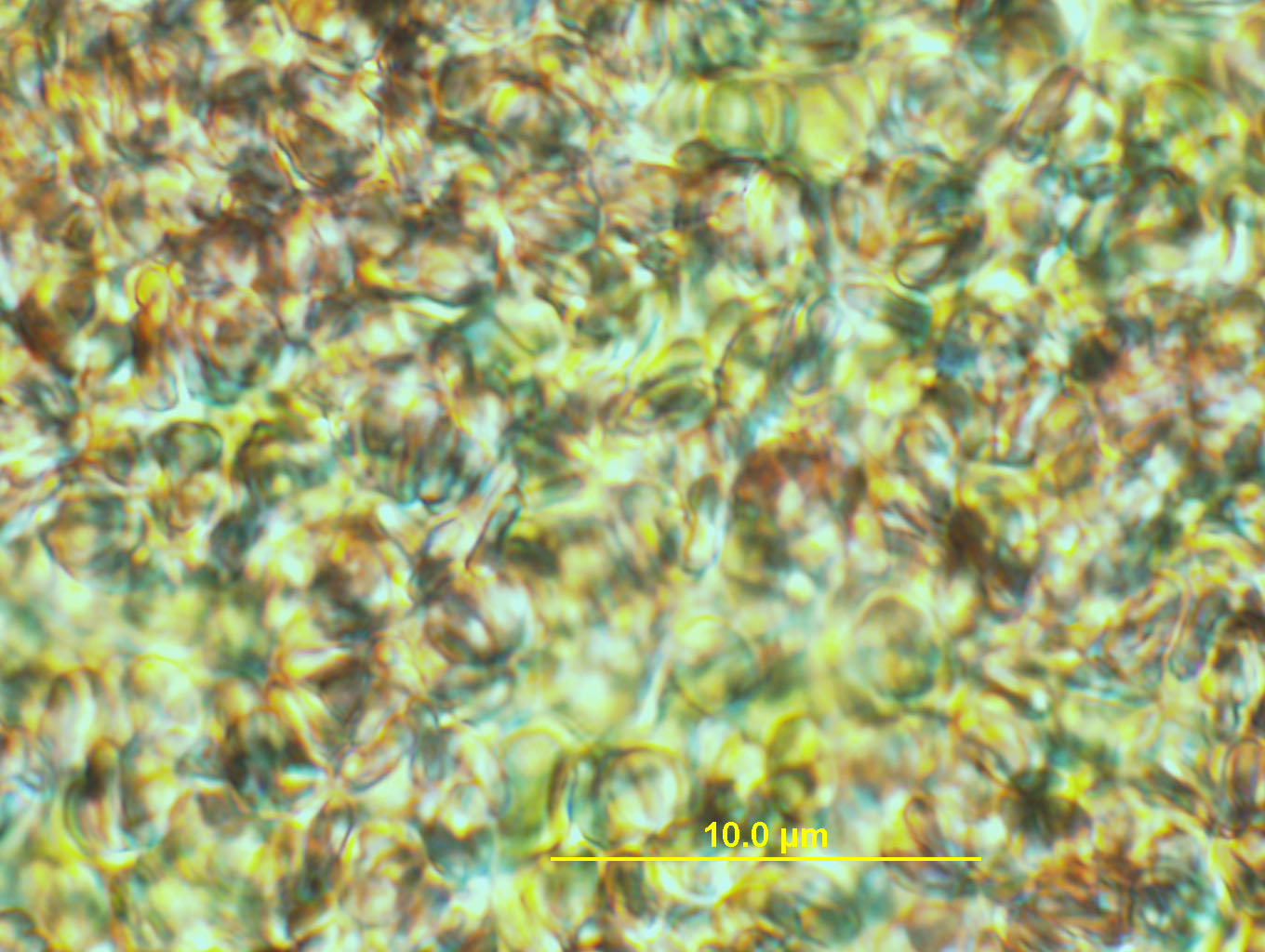
(above) human blood 40X objective white light
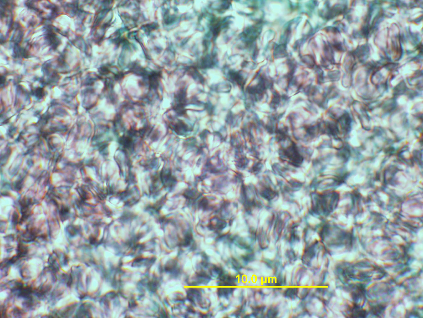
(above) Blood 20x with DIC (Nomarsky) filter (white light)
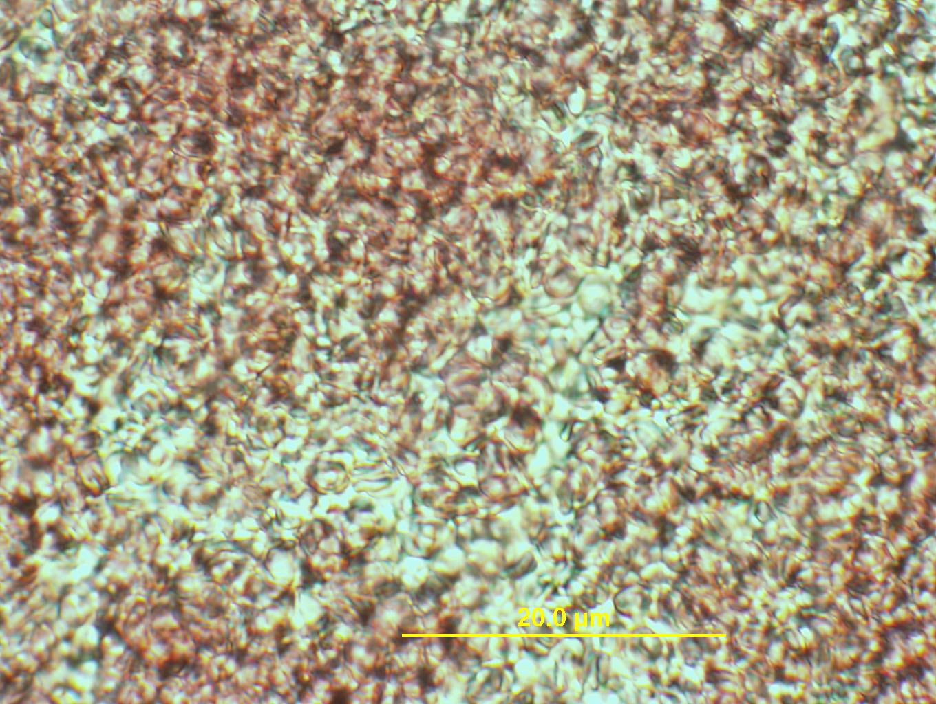
(above) Blood 40x with DIC (Nomarsky) filter (white light)
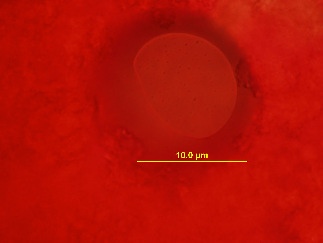
(above) Blood Cell with main red filter
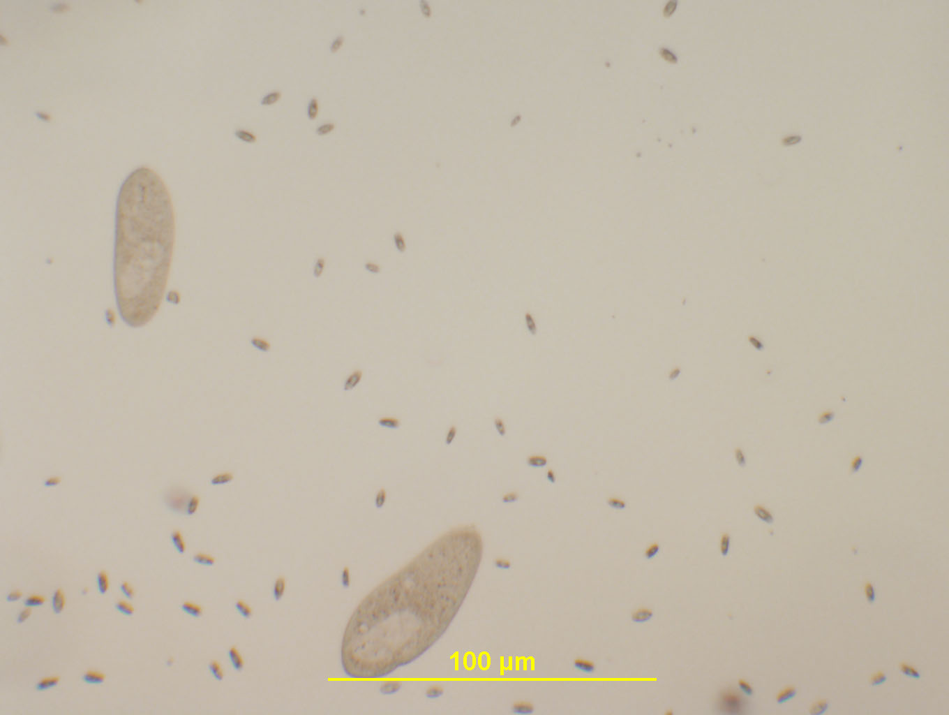
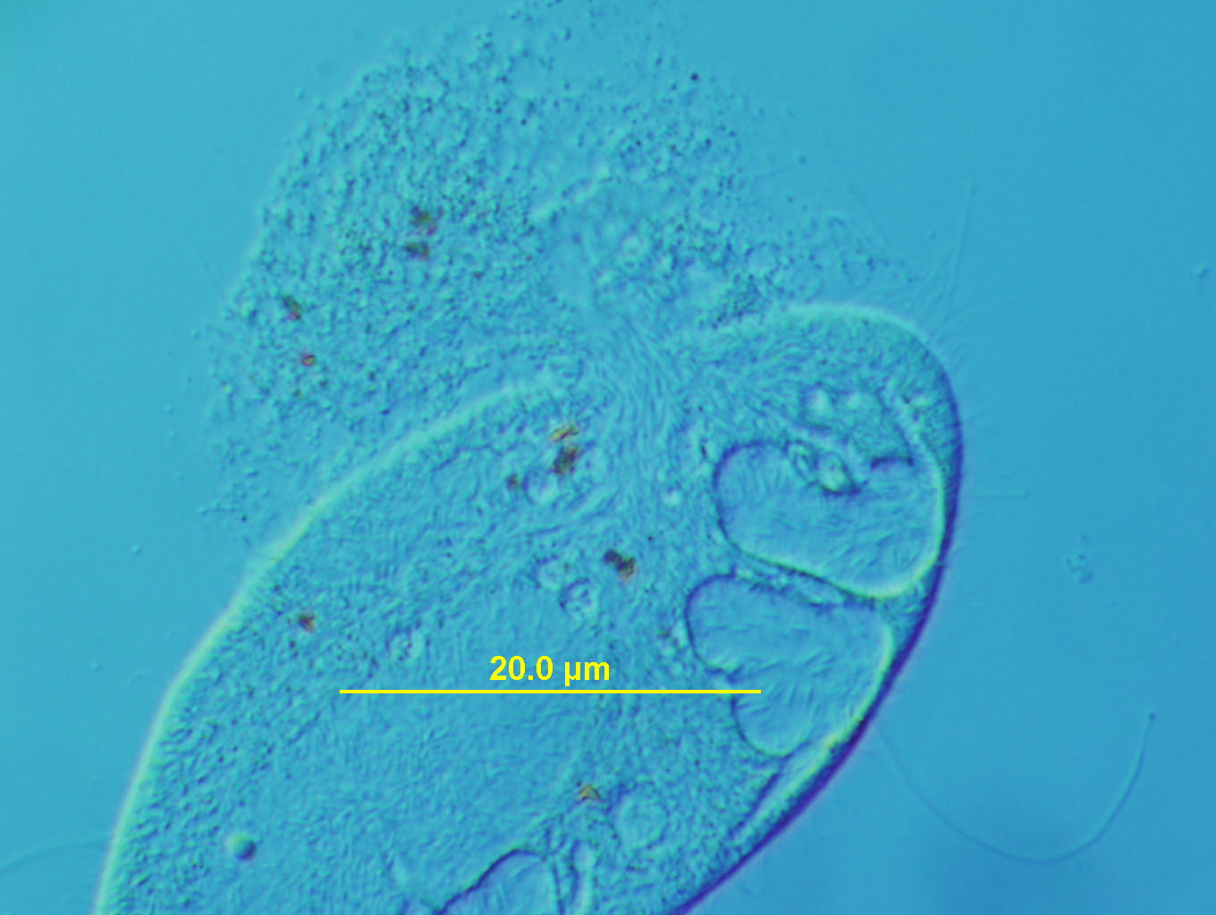
(above) Paramecium at 4X, white light...lots of small swimming "babies" around
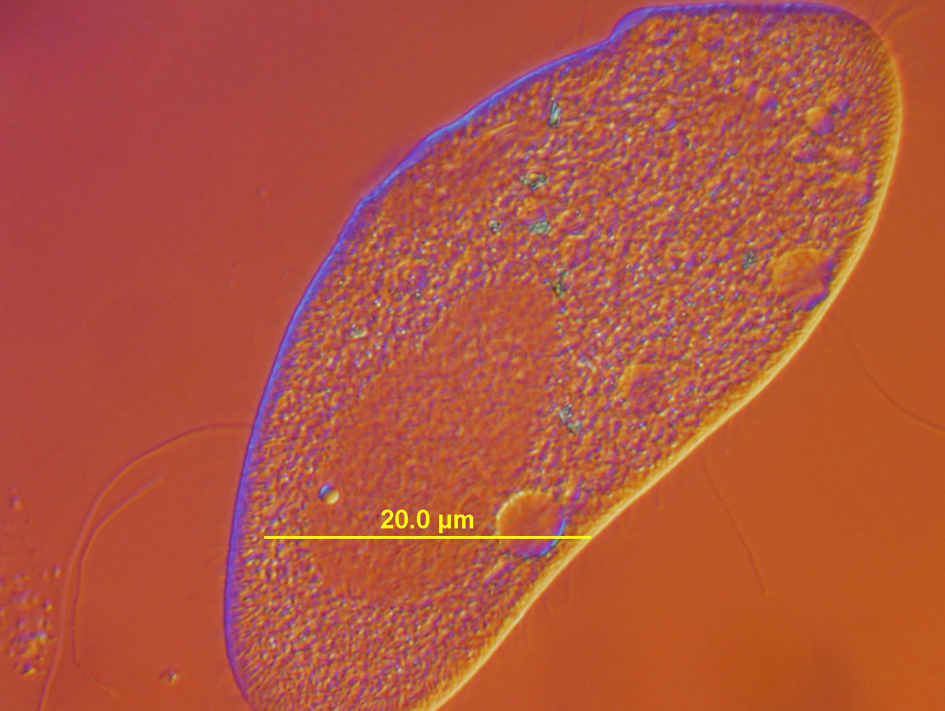
(above) Parameciums 10x objective, white light

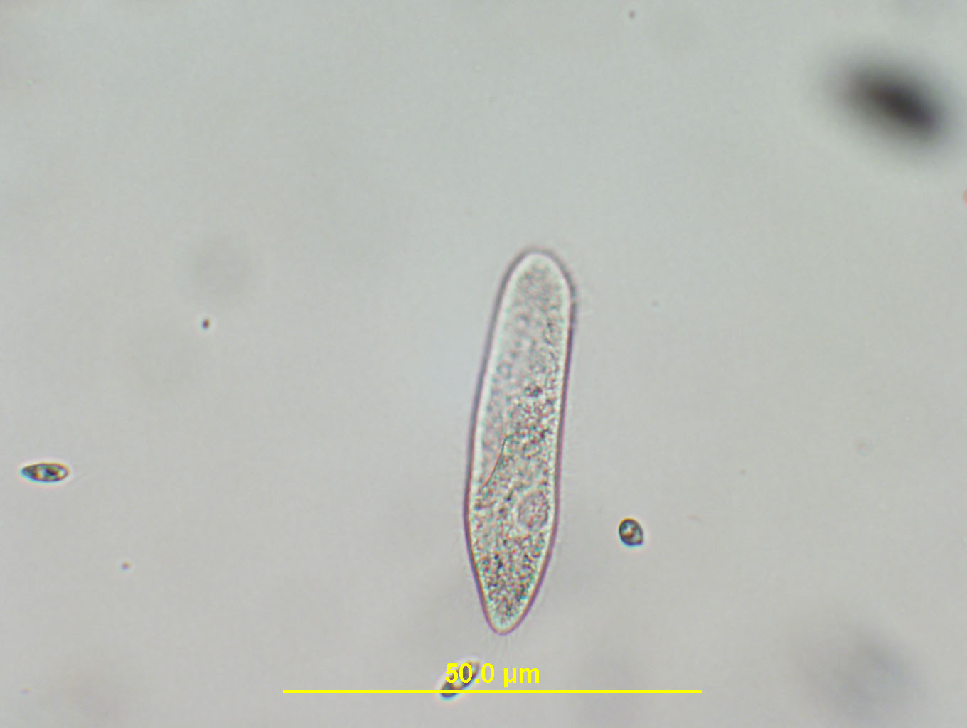
(above) Paramecium Caudatam, 10x, white light, DIC (Nomarsky) filter. This organism has been on the slide for about 45 minutes...at which point blisters begin to develop around the outside of the outer cell membrane. Shortly afterwards, the cell membrane bursts open and the contents of the organism spill out. Paramecia survive a maximum of about 1 hour on a slide after which they simply disintegrate leaving a formless remnant.
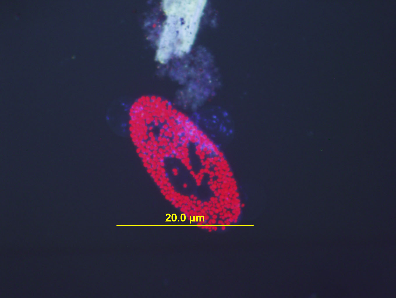
(above) Paramecium Bursaria at 20X under Ultraviolet light. These organisms, who have green spots under white light, must have cholorplasts in them as they flouresce under UV light. This organism has a symbiotic relationship with green alga called Zoochlorella who live inside the Paramecium and provide it with food. The paramecium provides the alga with protection and a means of "getting around". (Hey, give me a ride and I'll cook dinner!).
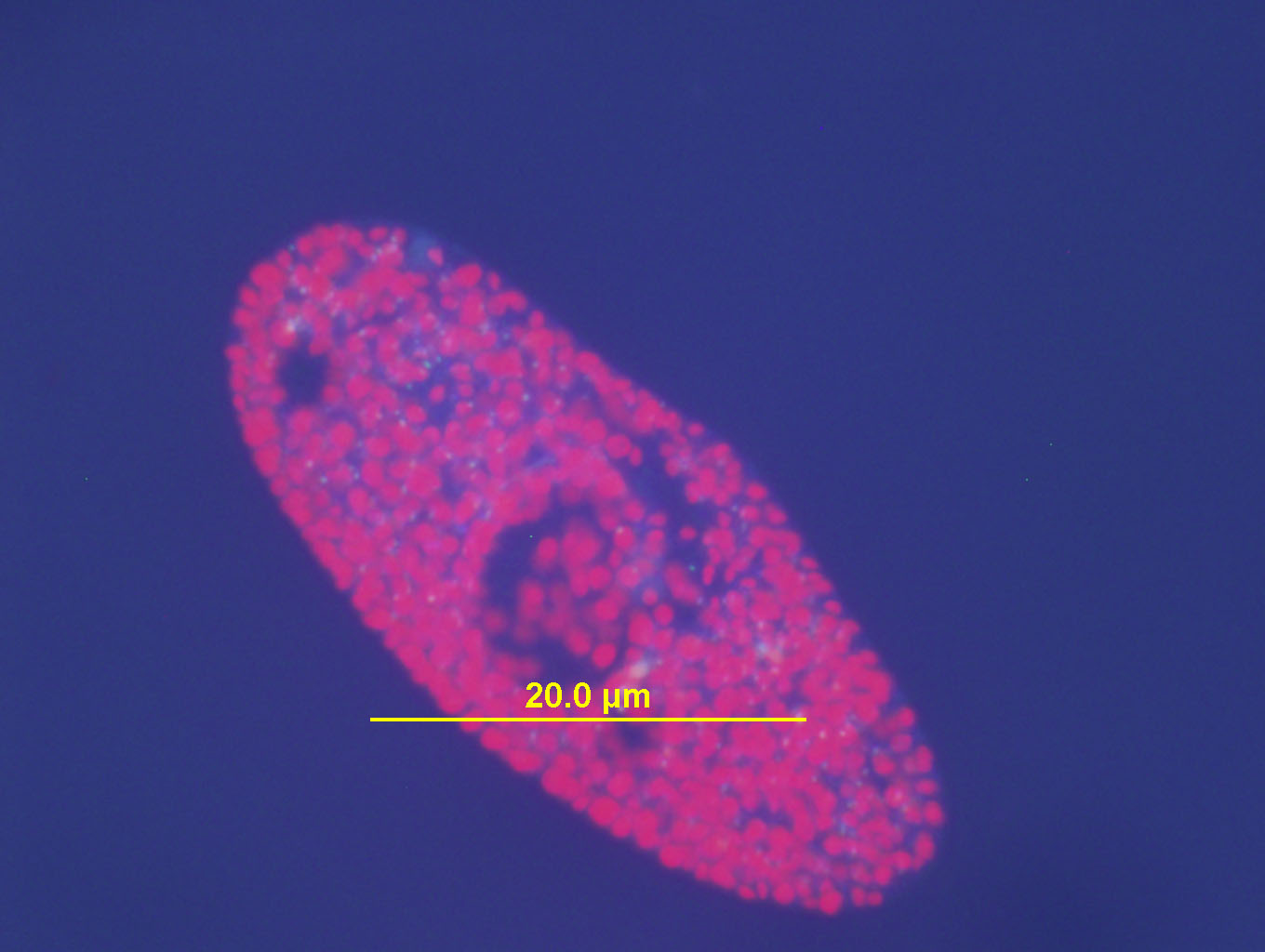
(above) Paramecium bursaria at 20X under UV light.
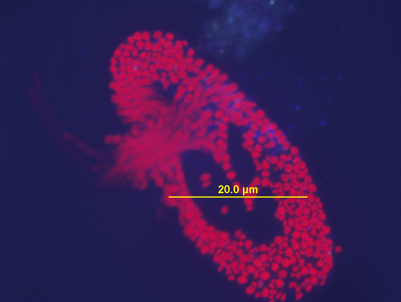
(above) Paramecium Bursaria 20X UV light: The cell wall/membrane has ruptured and the contents of the cell beging to spill out (upper left of organism). This is a natural occurrance. Nothing was done to try to destroy this cell. I was simply watching it over the course of an hour.
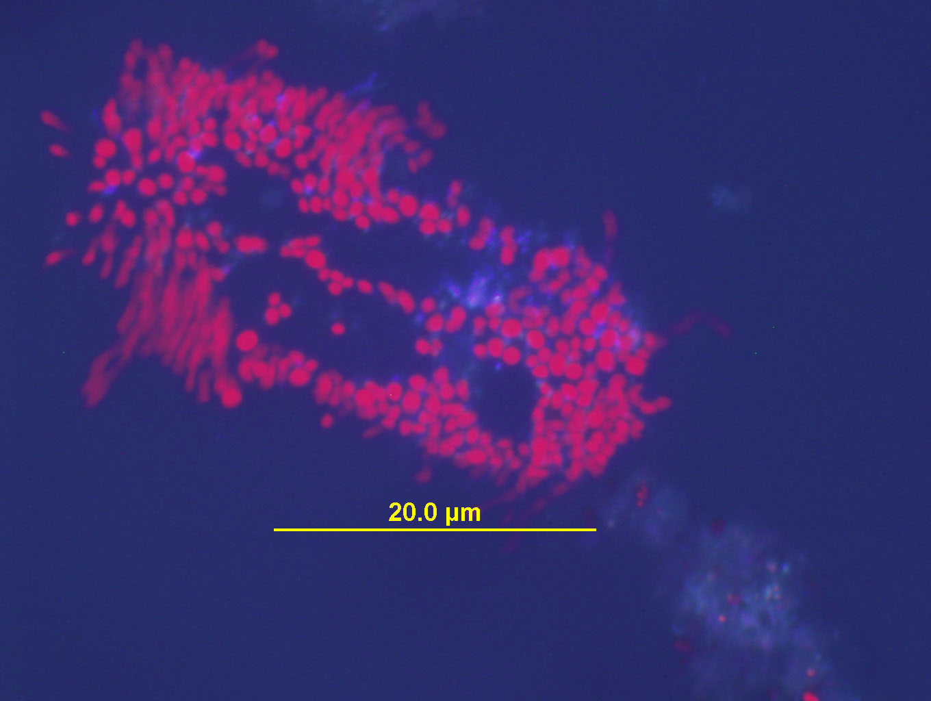
(above) the same Paramecium from above a short time later... starting to loose its form as it completely disintegrates (UV light).
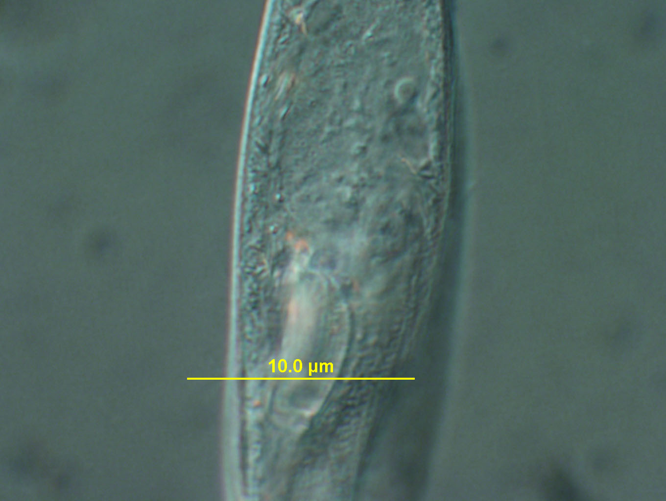
(above) Paramecium at 20X under white light with DIC (Nomarsky) filter. Note how the outer cell membrane has ruptured in the upper left corner of the organism...and the contents of the cell can clearly be seen spilling out of the organism. The Paramecium is in the process of dying after about 45 minutes on the slide. All Paramecium I have observed for an hour or more on a single slide will go through this process and eventually all die.
(above) Paramecium 20x DIC filter. The DIC filter bends light, casting different frequencies of light toward the organism. In some cases, certain wavelengths of light will cause different parts of the organism to flouresce or appear more brilliant.
(above) Paramecium 20X DIC filter with RED main filter added.

(above) Paramecium at 40X with DIC (Nomarsky) filter. Notice how some parts of the organism seem to flouresce with the DIC filter adjusted in a specific manner.
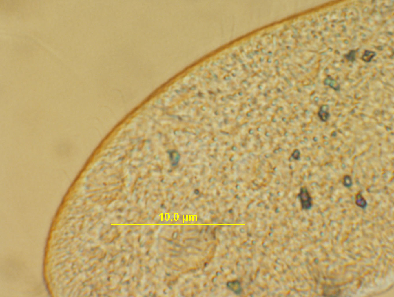
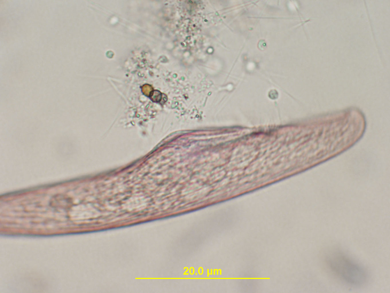
(above) Paramecium at 40X with DIC filter. Notice how the DIC filter can bring out various textures of the organism.
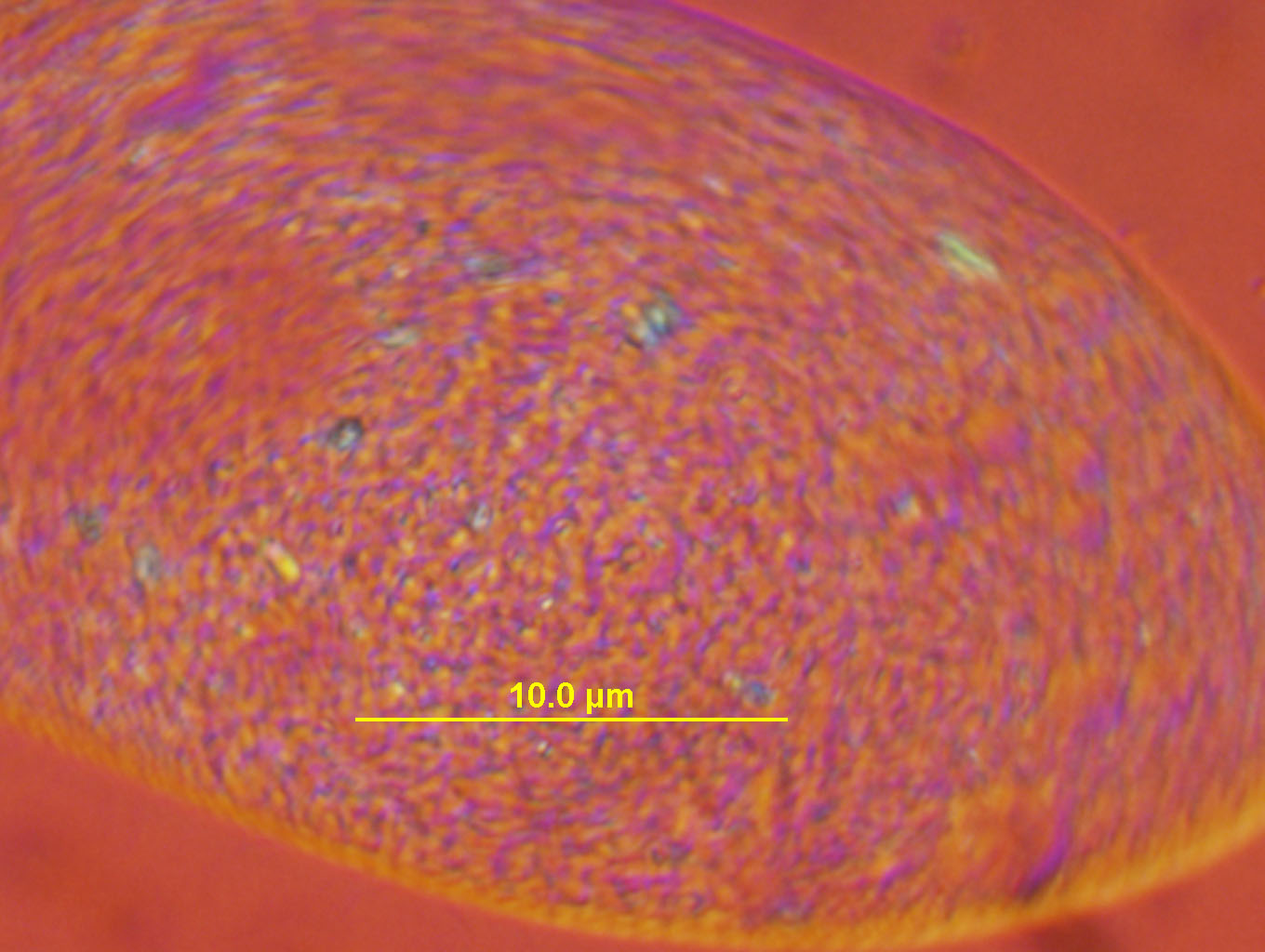
(above) A different setting of the DIC (Nomarsky) filter at 40X. Lovely color and a different set of details emerge.
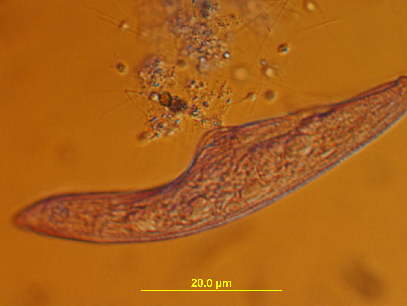
40X objective, DIC filter and RED main filter.

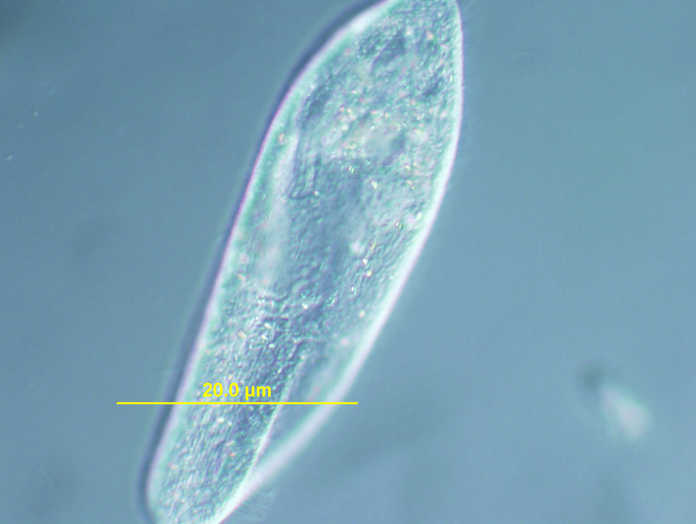
(above) Blepharisma at 10X under white light. Blepharisma are very difficult to photograph because they swim very fast across the slide. You have to search long and hard to find one that is "resting" and quickly take its photo. (Or you can use something called "Protoslo" which will slow them down). These photos of Blepharisma are the result of me chasing them on the slide for quite a long time (no Protoslo used ).
(above) Blepharisma at 20X under white light. Notice the "striations" along the body.
(above) Blepharisma at 20X under white light with the DIC (Nomarsky) filter. Even more detail of texture is made apparent with the DIC filter.