
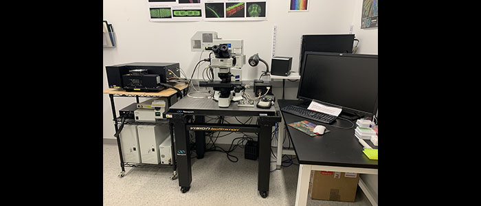
Olympus Fluoview 1000 confocal laser scanning microscope
Confocal Laser Scanning Microscopy Laboratory
SMIC maintains an Olympus Fluoview 1000 confocal scanning microscope supported by the funding from the National Science Foundation's Major Research Instrumentation Program.
Equipment
Olympus Fluoview 1000 confocal laser scanning microscope: this state-of-the-art confocal microscope is the latest addition to SMIC. For more information, see the Olympus brochure.
- Specifications:
- Spectral and filter scan units
- 5 laser channels
- High sensitivity GaAsP Detector for imaging low level fluorescence
- SIM Scanner for simultaneous photostimulation during time-lapse imaging
- Enhanced reliable objectives for live cell imaging
- High-precision focus and Z-tracking through extended time-lapse imaging
- 3D rendering and image analysis with advanced software
- Spectral unmixing for separating similar fluorophores
- Applications:
- Multi-dimensional time-lapse imaging of live cells/organisms
- High-resolution 3D rendering of Z-stack images
- FRET and FRAP technologies
- Multi-laser combiner for simultaneous imaging of up to five different fluorophores
- DIC imaging
- Multi-point, tile and mapping scans
- 3D Mosaic imaging