This is a series of microphotographs I took in June 2006 at the Skidmore College Imaging Center (SMIC) The photos were taken using an Olympus BX60 trinocular microscope with an Olympus DP70 digital camera attached to a computer. This microscope has many wonderful capabilities including two different light sources: tungsten (white light) and mercury light (which emits numerous powerful wavelengths includling blue, green and ultraviolet), plus the scope has a "DIC" filter which allows for some great variations in depth of field and color while using the white light source. **Special Thanks go to Professor David Domozych, Department of Biology at Skidmore College, for his generous assistance in learning how to make these beautiful microphotographs**. (note: all images are ©2006 Anthony G. Holland)
These are all microphotographs of a single celled organism: Gree Algae: Micrasterias
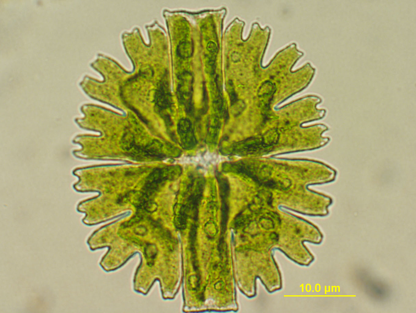
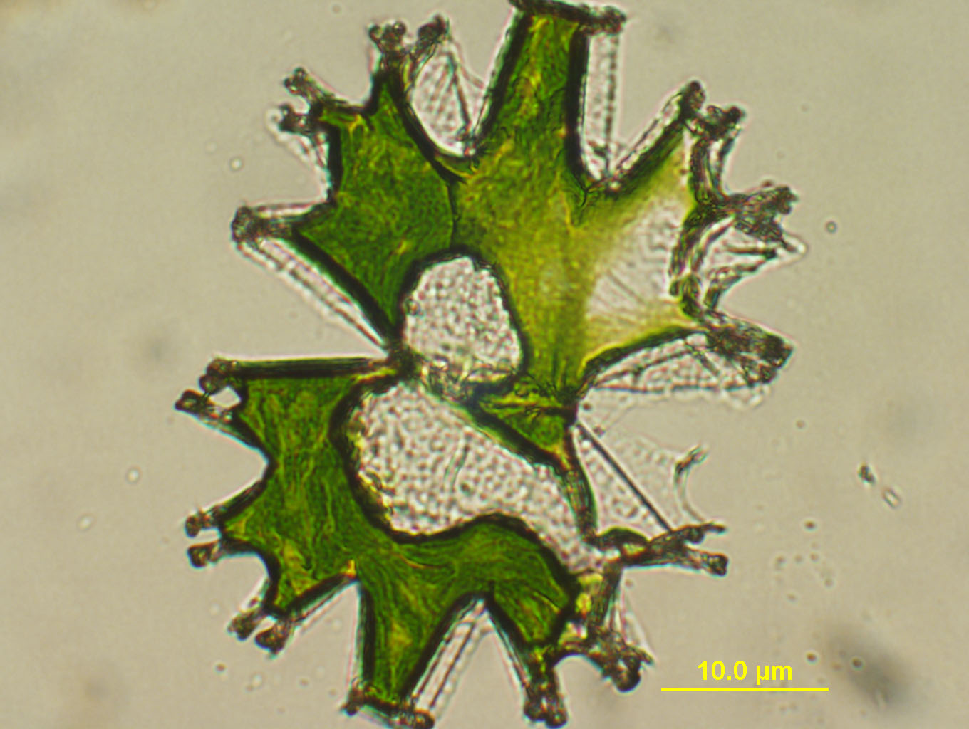
A marvelously beautiful organism! Above: white light, 20X objective.
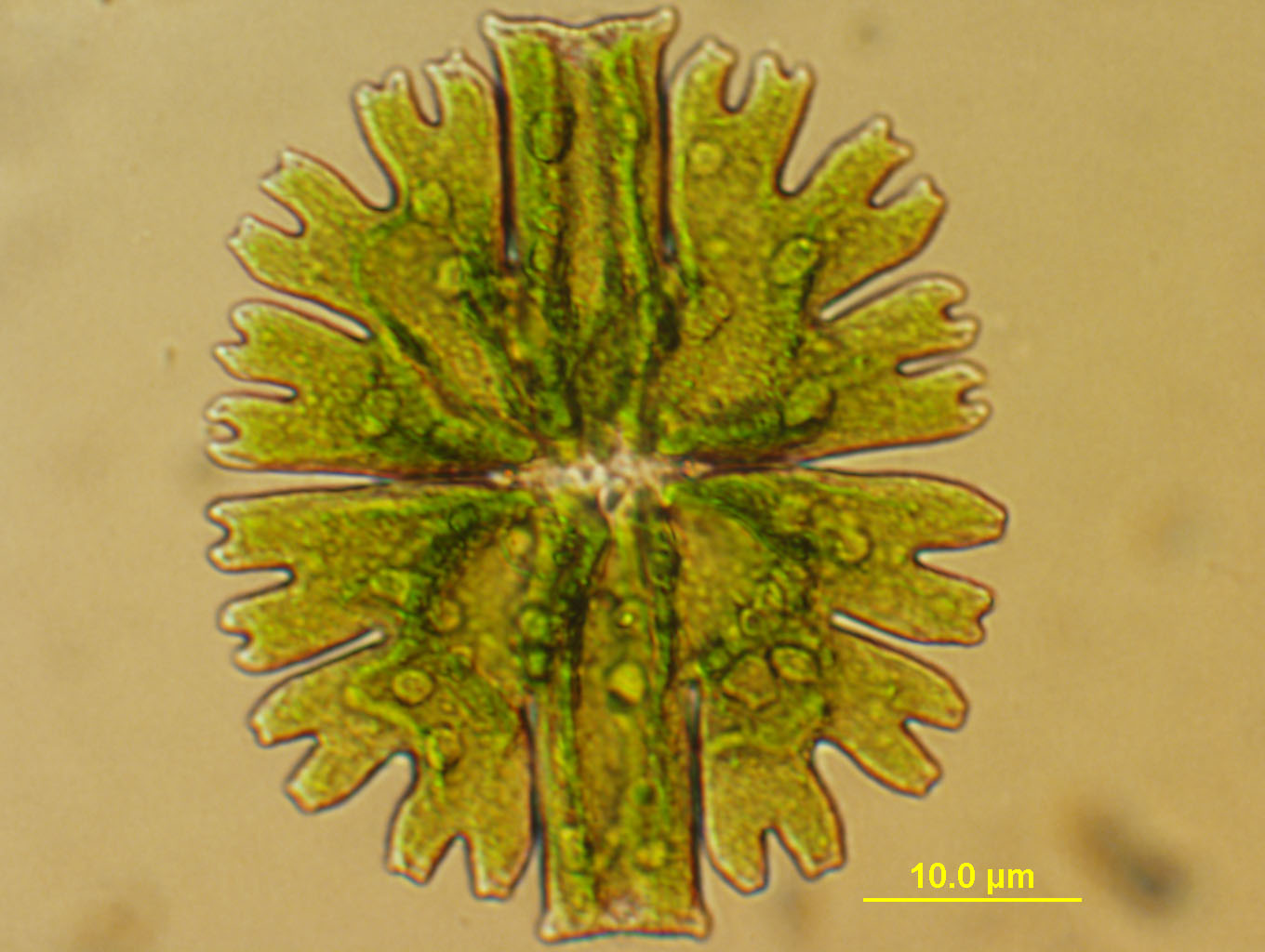
Above: white light, 40X objective. HIGHLY INTERESTING: tiny "eggs" were shooting out of the organism while I was preparing this photograph . In this photo, I managed to capture several small "eggs" recently ejected. Look at the center of the organism, then follow a line to the right... and you will see the path from which the objects were "ejected". It was remarkable live under the microscope, the velocity at which the small spheres are ejected was startling!
Above: White light but using the "DIC" filter. This gives variations in color, depth of field and level of resultion and detail.
Above: white light again with the DIC filter, different filter setting (20X objective).
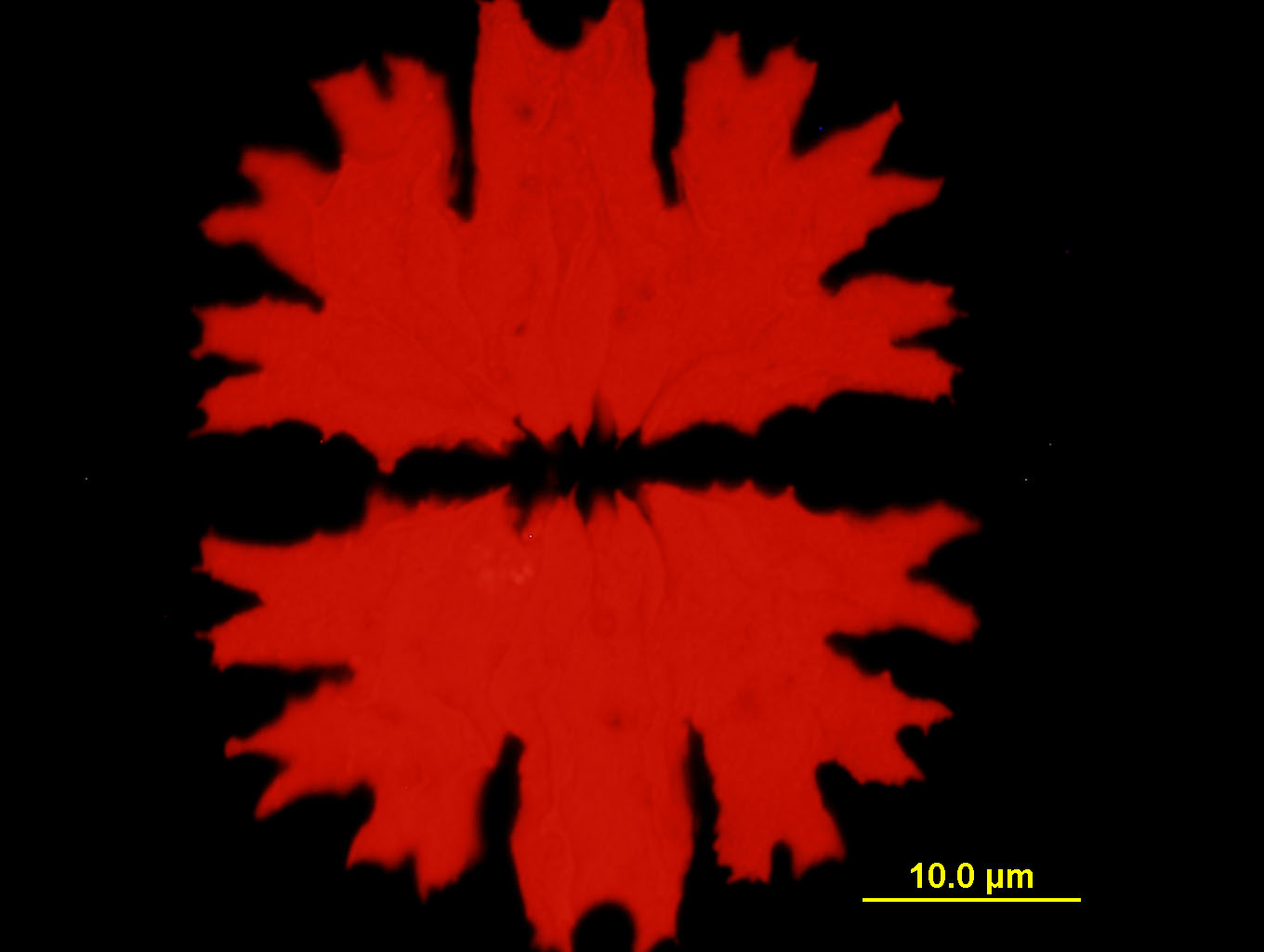
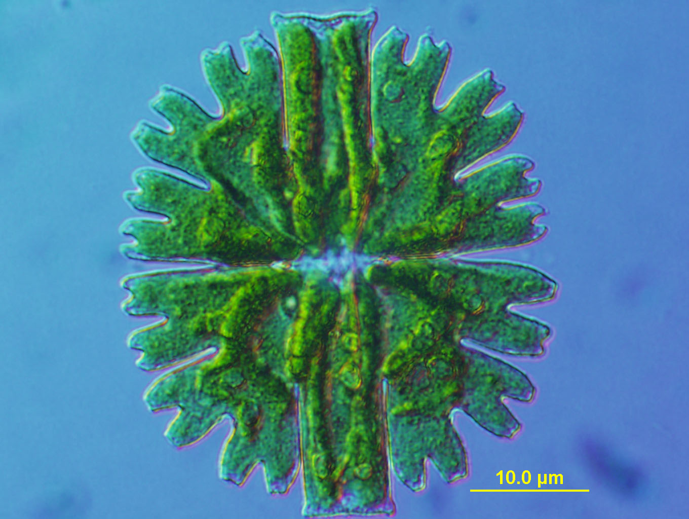
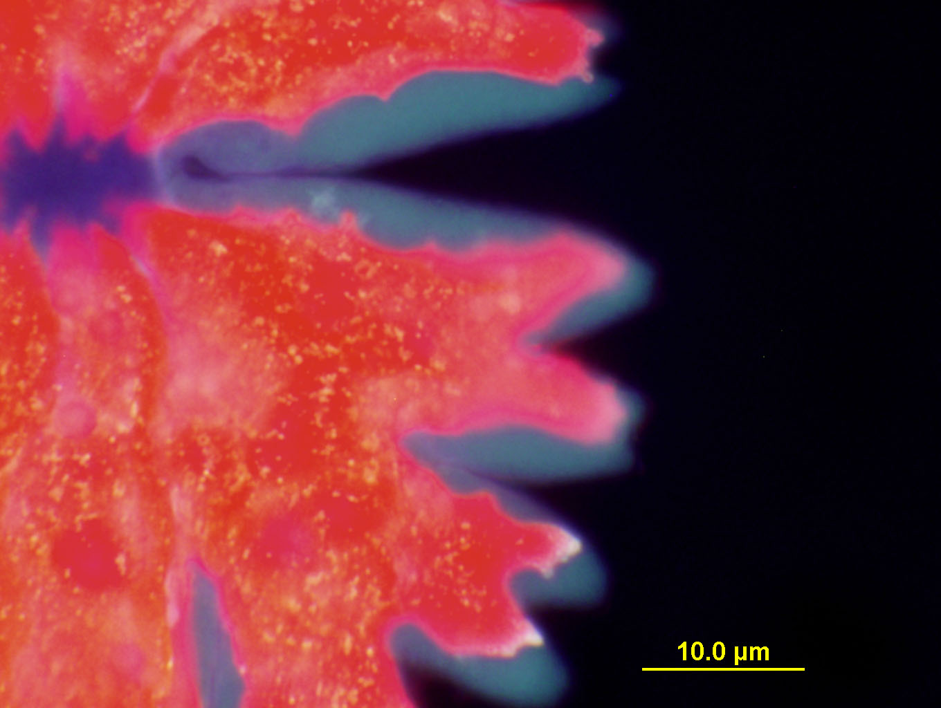
Above: Mercury light with BLUE filter: causes flouresence in red. Green Algae contain lots of clorophyl and convert sunlight into energy for the plant. Clorophyl will naturally flouresce under the intense blue light from the mercury lamp without any additional dyes or markers needed. Absolutely marvelous colors!
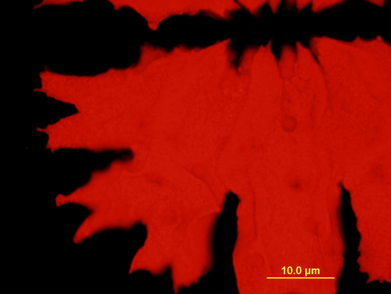
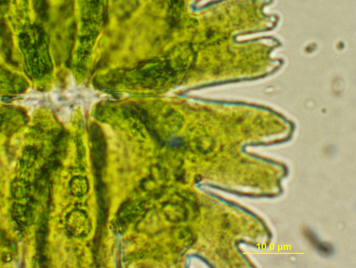
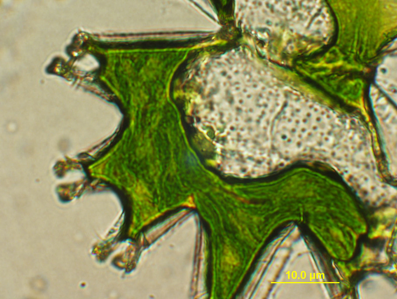
Above: again with the mercury lamp but with the 40X objective. I believe the scale marking here is off as I forgot to tell the computer that I had changed the objective! Sorry about that! I'll fix that next time around! In this photo, I was going for details along the left edge of the organism. There are some very interesting shapes along the left "leaf-like" edges.
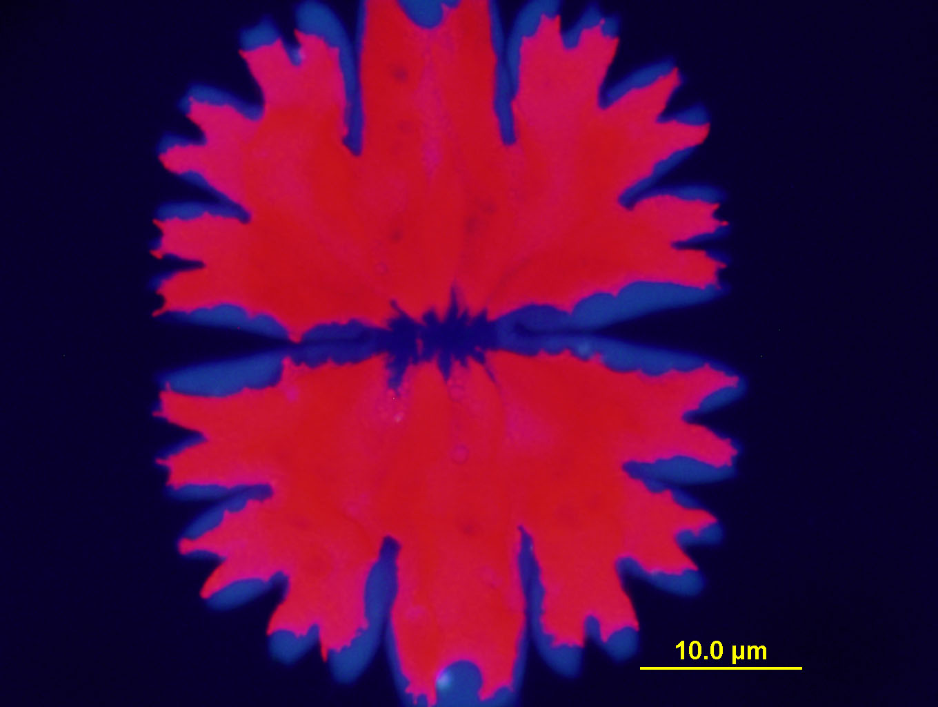
Above: Ultraviolet Light flourescence! After much reassurance from Professor Domozych that I would not blind myself with the UV light source coming from the mercury lamp, I boldly shot this photo. I just love the capabilities of UV light.
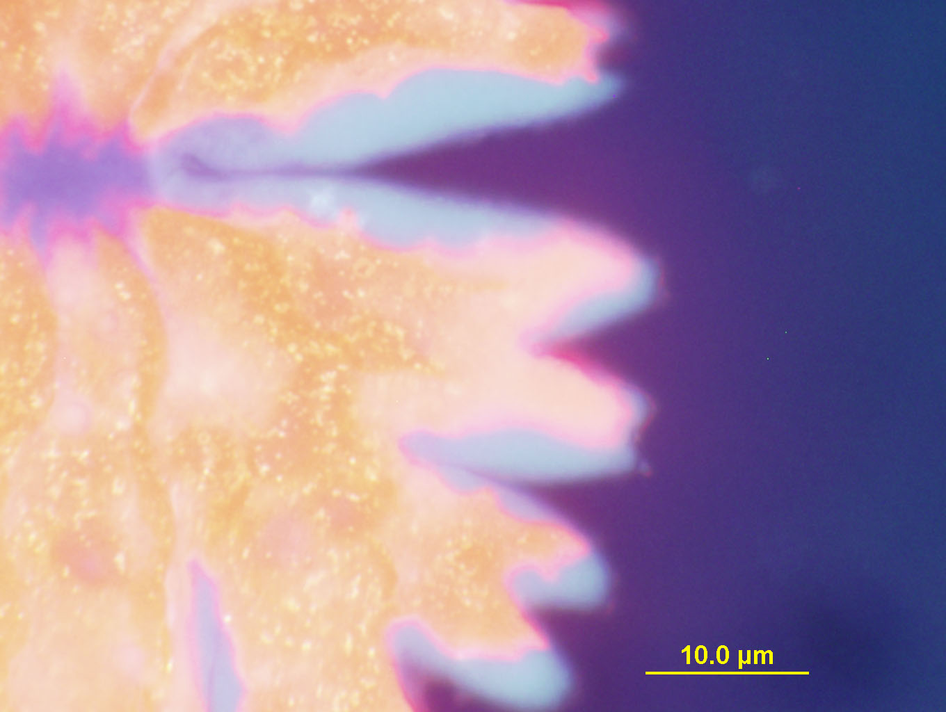
Above: 40X objective and a different setting with UltraViolet Light. The scale is off here unfortunately, but I love the colors that the UV brought out in this exposure.
Above: 40X objetive with UV light again but with yet another setting on the exposure time etc. on the computer. I just love this photograph. I think the colors are really magnificant. I love the details of the bright spots, the white tips and the aura-like glow along the outer edges. For me, this image evokes music in my composer "inner ear". I am inspired to try and think of a new musical composition which will evoke similar feelings using music. This image speaks to me in a music which is delicate, distant, faint.
Above: I'm not sure what this is. In my notes, I call it "broken Algae". Perhaps it has not completed its formation? Perhaps it was damaged when I was moving it from its container to the slide? The scale here is correct with the 20X objective.
Above: "broken Algae" again but with the 40X objective. There is a very interesting pattern in the series of "dots" inside the core of the organism...and that core is so much larger than that in the organism in most of the other photos above. Interesting also are some "dots" that appear to the left of this organism.... some of these "dots" were swimming madly in clockwise circles but I did not have a powerful enough objective to see the morphology of those madly swimming dots. When first I was learning to use the microscope to take these photos, the organism "moved" on the screen... and I was startled! It was alive! Imagine what wonders we could see as we enter an ever smaller world, view it, photograph it, visit it and marvel at the biological constructions on our earth! What a joy it was for me to learn how to take these wonderful photos. Many thanks to Professor Domozych!!!