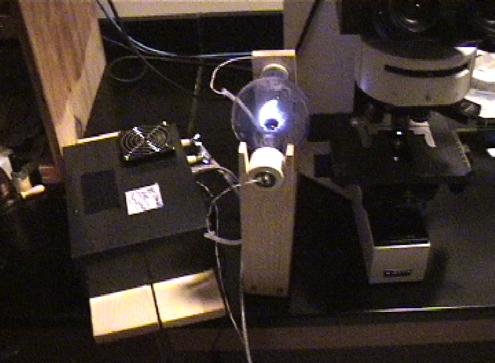
This series of microphotographs and video was shot in the Skidmore College Imaging Center (SMIC) The images were taken using an Olympus BX60 trinocular microscope with an Olympus DP70 digital camera attached to a computer. This microscope has two different light sources: tungsten (white light) and mercury light(UV), plus the scope has a "DIC" filter. **Special Thanks go to Professor David Domozych, Department of Biology at Skidmore College, for his generous assistance**.(note: all images are ©2007 Anthony G. Holland)
----------------------------------------------------------LOG ENTRY FOR 8/27/07 (These are the most dramatic results I've obtained so far in these experiments)---------------------
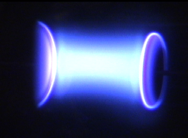
After a short summer break and the installation of a new amplifier (100 Watt Class C to which I added a small cooling fan), the results of this night's experiments were absolutely outstanding! More than 20 organisms were filmed LIVE while disintegrating/exploding under the influence of the Plasma light, and many more were 'devitalized/disabled/immobilized'. I had to remind myself.... we are seeing these dramatic results using only a 100 watt amplifier and with the plasma bulb (phanotron) about 5 inches away from the microscope slide!
CLICK ON THE PHOTO BELOW TO SEE THE LIVE VIDEO FROM THE 8/27/08 EXPERIMENT (BEST SO FAR!)
pictured above: a 'double' disintegration/explosion of two organisms occuring simultaneously (very rare to catch on video!)
CLICK HERE TO SEE STILL PHOTOS TAKEN FROM THIS LIVE VIDEO
| Click Here to Go to Most Recent Experiments (or continue below to see some 'historical' experiments) |
-----------------------------------------------------------LOG ENTRY FOR 7/7/07 (another lucky day in the lab!)--------------------------------
Once again had some great results in the lab tonight, including some rather 'dramatic' "devitalizations" caught on live video. I seem to be getting rather consistent results now in using the Plasma beam to disintegrate these small organisms. Click on the Photo below to see the VIDEO HIGHLIGHTS from 7-7-7.
| OR CLICK HERE TO SEE PHOTOS TAKEN FROM THE LIVE VIDEO |
CLICK ON THE PHOTO BELOW TO SEE THE LIVE VIDEO FROM THE 7/7/07 EXPERIMENT
------------------------------------------------------------LOG ENTRY FOR 7/6/07--------------------------------------------------------------------------
Had some dramatic results this night in the lab. Click on the Photo below to see the video highlights on google video
Click on the photo above to see video highlights of the July 6, '07 plasma experiment, OR CLICK HERE TO SEE ONLY PHOTOS
--------------------------------------------------------------------------------------------------------------------------------------------------------------------------
| Continue below or Click Here to see more dramatic video and photos of Rife-Bare Plasma Experiments on microorganisms (July 16, 2007 experiments and later) |
| CLICK HERE to learn about a FREE PROGRAM ("RBSQuareGen" for Macintosh Computers only) which was used to create the effects you see in these plasma experiment videos (includes free download link) |
| CLICK HERE to see waveforms and spectral analysis of the frequencies used to cause the dramatic effects seen in the videos of these plasma experiments |
-----------------------------------------------------------------LOG ENTRY FOR 6/28/07----------------------------------------------------------------------
Thursday, June 28, 2007
This was an historic experiment. I slightly altered the program on my F125 frequency generator (programming code to be listed below soon) and had some amazing results. For the first time in months of experiments, many many organisms were either immobilized/devitalized or totally disintegrated/destroyed. It was a night for dramatic observations and luckily I had my video camera with me. Click on the photo beow to see a video of the live experiment (it's a Google Video link):
CLICK ON THE PHOTO ABOVE TO SEE REAL TIME VIDEO OF THIS HISTORIC EXPERIMENT OF JUNE 28, 2007
---------------------------------------------------------------------LOG ENTRY FOR 6/25/07--------------------------------------------------------------------
---------------------------------------------------------------------------------------------------------------------------------------------------------------------------
Monday, June 25, 2007
This night I had some success video taping the destruction ('devitalization') of a Blepharisma which I believe was caused by my Rife-Bare plasma device running a custom series of frequencies I designed for the F125 Frequency generator. While there are still many more variables to be understood in this process, I believe this video documents a 'recapturing' of the effects described by Royal R. Rife in the 1930's. This video segment runs on Google Video and is a real time video of the relative success of this night's experiment (click on photo below to see video):
CLICK ON THE PHOTO ABOVE TO SEE THE VIDEO OF THIS EXPERIMENT
---------------------------------------------------------------------LOG ENTRY FOR 6/22/07---------------------------------------------------------------------
Friday, June 22, 2007
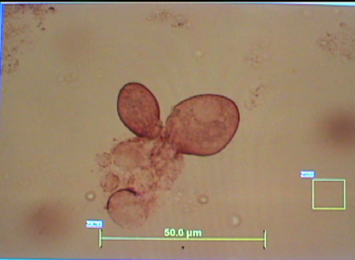
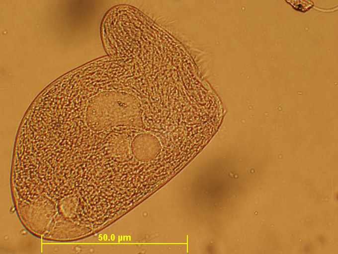
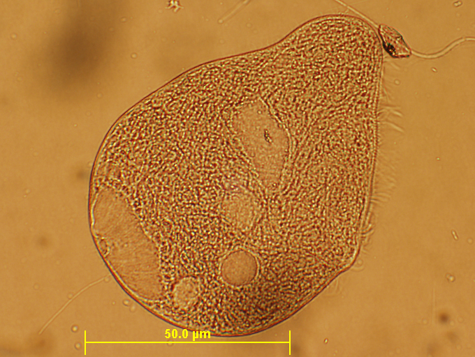
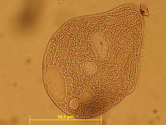
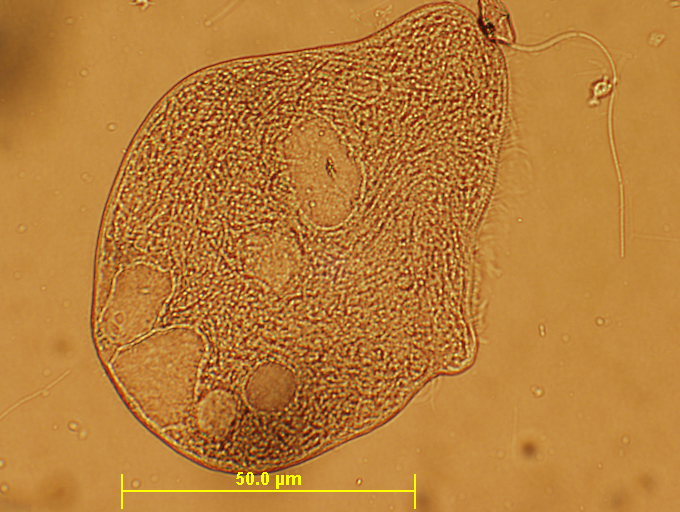
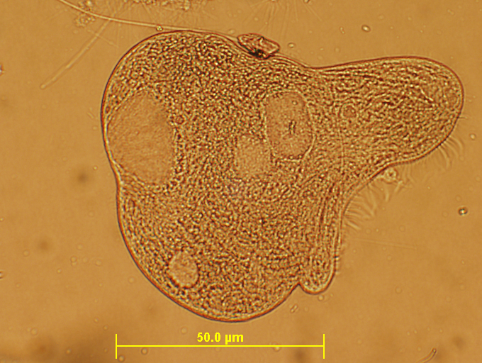
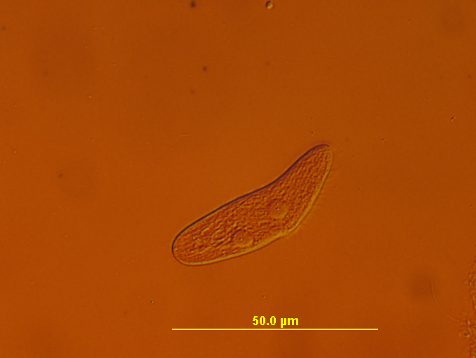
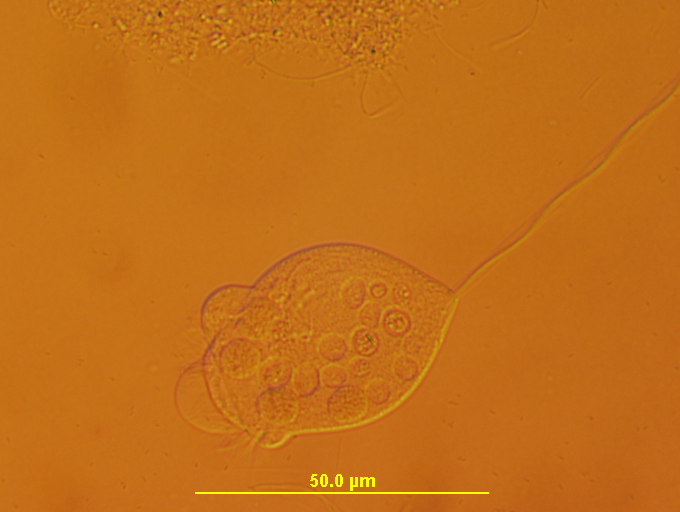
Seem to have managed to duplicate the results from Feb. 18 2007 this night. The key seems to be a program I developed on the F125 frequency generator which uses a second RF carrier (called a 'backfreq' in the F125 programming). When this second carrier is approximately 11X the main frequency for the Blepharisma (currently operating under the advice that 924 is a 'main' freuqency for Blepharisma), we seem to find at least one organism on the slide which is almost immediately affected by the plasma. The organism quickly stops all movement and over the course of the next 27 minutes becomes terribly mishapen with internal structures seeming to disintegrate. The original shape and internal structuring of the organism is never regained after running the experiment and the organism never swims again. We continue to see cilia movement for some time afterwards but the organism is not capable of any directed movement. I believe its normal biological functions are completely distrupted, meaning it cannot 'feed' in any way, cannot grow, cannot reproduce and cannot perform any of its normal biological functions. Royal Rife might call this organism 'devitalized'. For all practical purposes, the organism is rendered helpless and 'deactivated'. Were this a pathogenic organism, it's ability to cause disease might be ended.
A series of time lapse photos taken over a period of about 20 minutes. Note that the initial effects were seen almost immediately upon starting the experiment. Further refinement of this experiment is needed in order to determine with greater precision the most effective frequencies and the shortest amount of time possible to achive maximum 'deactivation' of the organism. Questions still remain, including: why all such organisms on the same slide were not so affected; if there is a relationship between the geomotry of the plasma bulb's electrodes and the effect induced on this organism, if the plasma wave is producing a directed scalar wave and numerous other questions remain. That being said, some level of success is seen here and in the February '07 experiments:
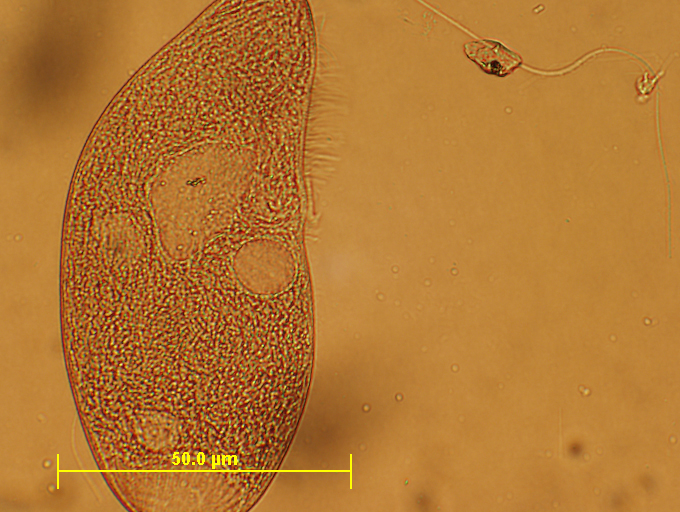
In the photo above, the Blepharisma has stopped swimming. This is usually the first sign that you have hit upon a 'sensitive' frequency for the organism. The microscope objecive is 10X here.
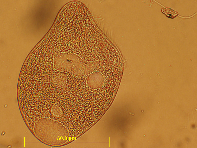
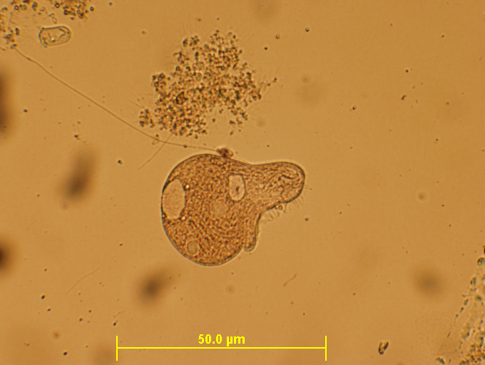
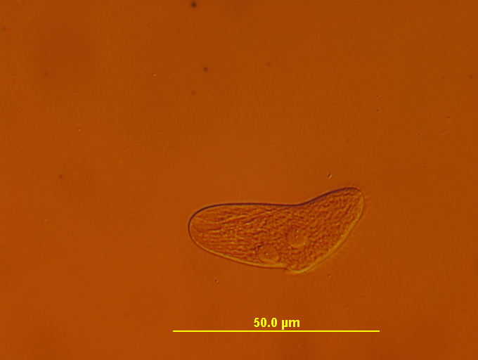
Within a few minutes, we see the organism (above) starts to become distended. This is the second step.
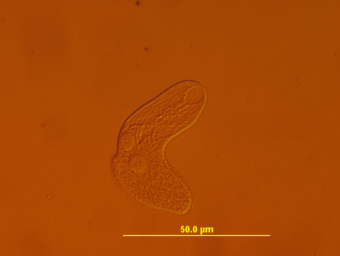
The organism continues to become further distended, gradually becoming more round than oblong.
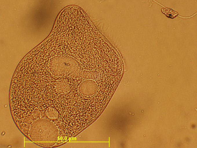
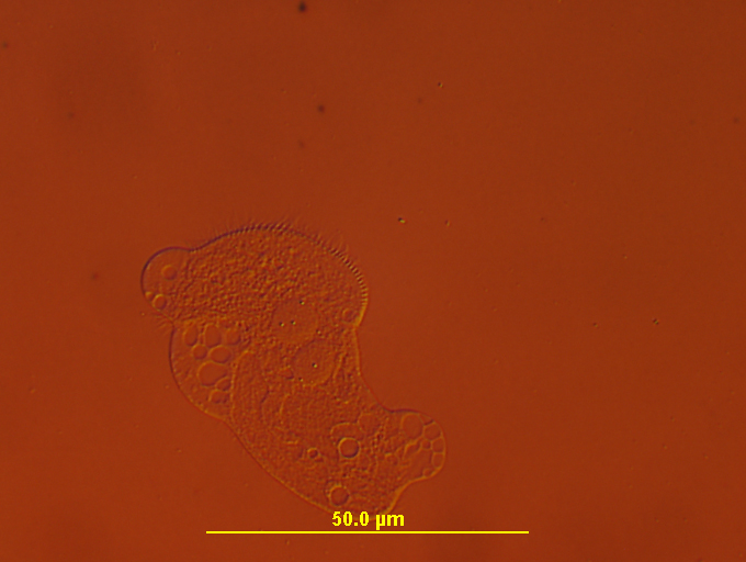
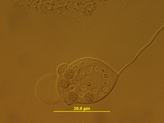
In the photo above, it seems the organism makes one last attempt at directed movement, but it can no longer swim.
The organism (above) returns back to a progressively rounder shape.
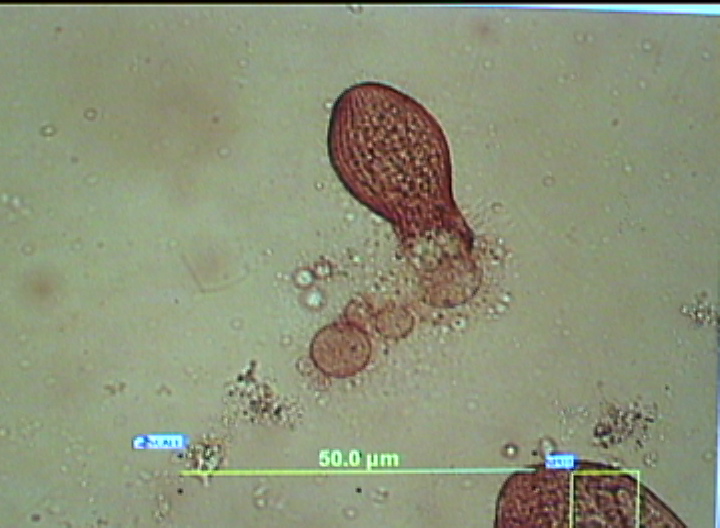
compare the internal structures of this photo (above) to the previos photo. Here you see a dramatic change in the internal structures and shapes.
Now we see (above) a small protuberance in the right side of the organism, while the internal structures continue to undergo change.
A dramatic shift (above) in the internal structures (study the left side of the photo and compare to previous photos) Note the addition of numerous internal 'spheroid' structures. This seems to be a characteristic of these plasma experiments on microorganisms.
Another shift in internal structures (above)
Yet another variation seen in internal structures (all these changes occuring within minutes of one another).
A new pattern emerges in the internal structures (above). Protuberance (bottom right of photo above) becomes more distinct/larger.
Final photo (above) with 10X objective. Milder cilia motion (right) persists despite no normal swimming motion from organism for over 20 minutes.
Final photo of organism with 4X objective. No swimming motion for 27 minutes. Badly mishapen. Internal structure disrupted. Organism is 'devitalized'.
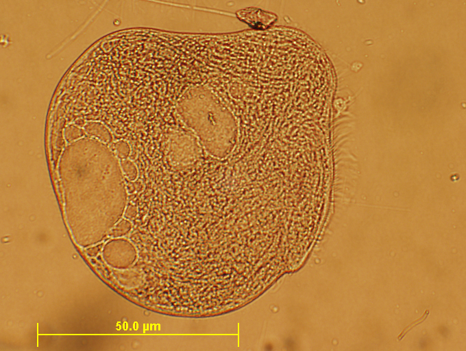
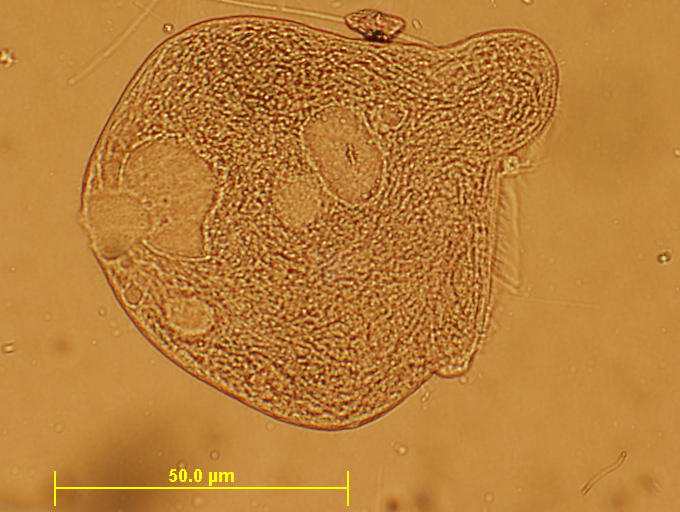
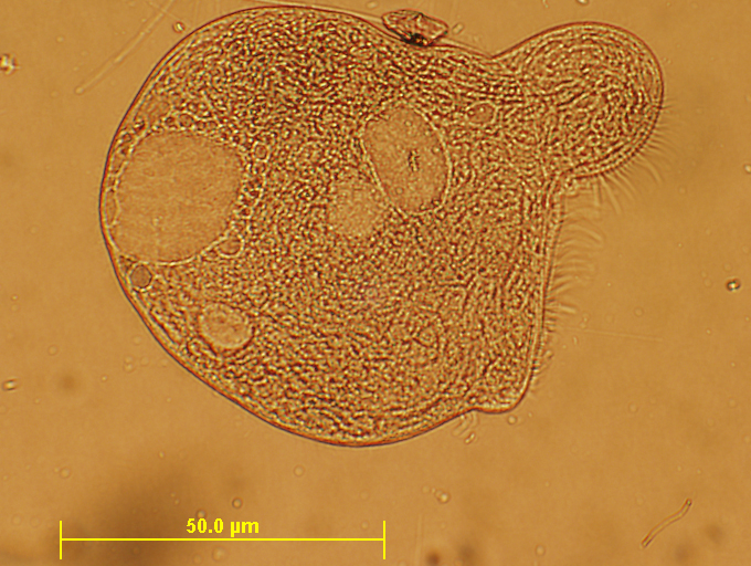
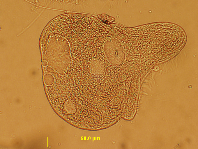
------------------------------------------------------------------------LOG ENTRY FOR 2/18/07-------------------------------------------------------------------
Sunday, Feb. 18, 2007
Here are a series of 'time lapse' photos taken during this experiment. Some success was seen in 'devitalizing' some Vorticella and also some Blepharisma. The Vorticella experiment ran a program on my frequency generator which I named "Vorticella10", which uses a second carrier wave whose frequency is approximately 10X the value of the audio range square waves being used to amplitude modulate the RF signal which is fed into the Phanotron plasma tube.
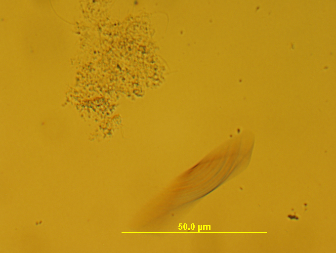
Normally a fast swimmer, the organisms above is just a blur to the camera (center right is the Blepharisma location)
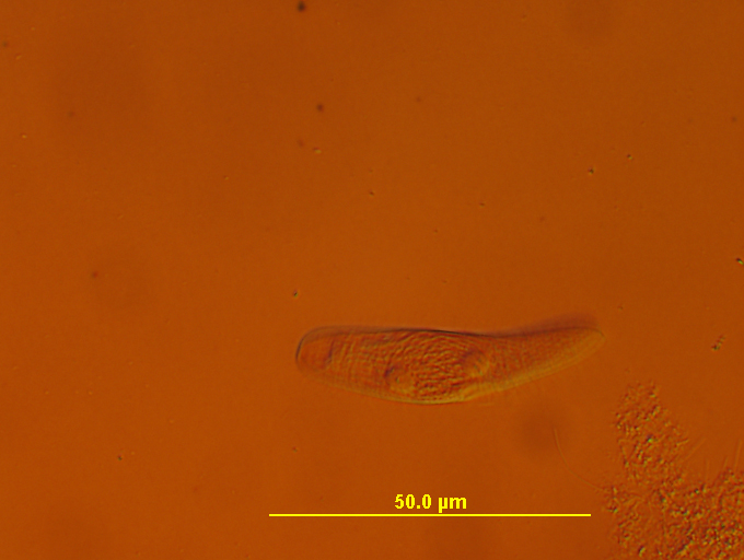
Above, the organism has slowed its swimming actions.... so we can take a better photo. Notice the organization and appearance of the internal structures become more apparent now (watch this carefully in the next few photos).
Above, the organisms has stopped swimming completely (no blur in photo) and along the right edge (center) begins to show signs of distension
Above, the organisms now showing signs of serious distension of bottom center edge and also grows a bit 'rounder' (length diminishes).
Above, same organism a few minutes later.... showing serious signs of malformation. Internal structures beginning to loose order.

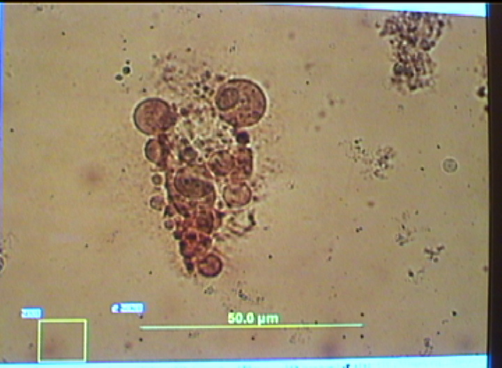
Above: same organisms now showing considerable internal disintegration and mishapen body.
Final photo (above) taken about 30 minutes into my computer program "Blepharisma10" on the F125 frequency generator. Outer cell membrane completely distorted. Internal structures nearly completely disintegrated. Note the development of large holes or 'spheroid' structures internally. This organism is now 'devitalized': it cannot maintain its normal biological functions.
Some photos of 'devitalized' Vorticella. "Devitalized means": no movement of any kind following the 30 minute treatment. No cilia action, no contractions of the stem. Sometimes small 'blisters' form near the opening of the organisms, possibly indicating a cell membrane rupture somehwere. Sometimes the organism swells into a round shape or develops an increased number of 'spheroid' shapes internally. (note: I believe the scale is incorrect in these photos, unfortnately.... forgot to tell the comptuer which objective was on the microscope at the time).
ˆ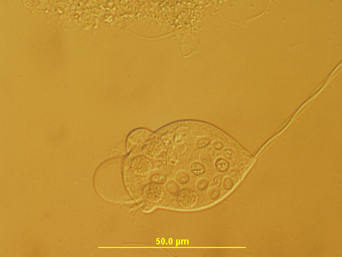
(10X objective with DIC filter)
note increase in size of blister in devitalized Vorticella (above)
Scale is correct in the photo above.