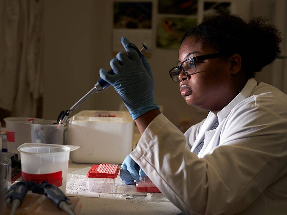
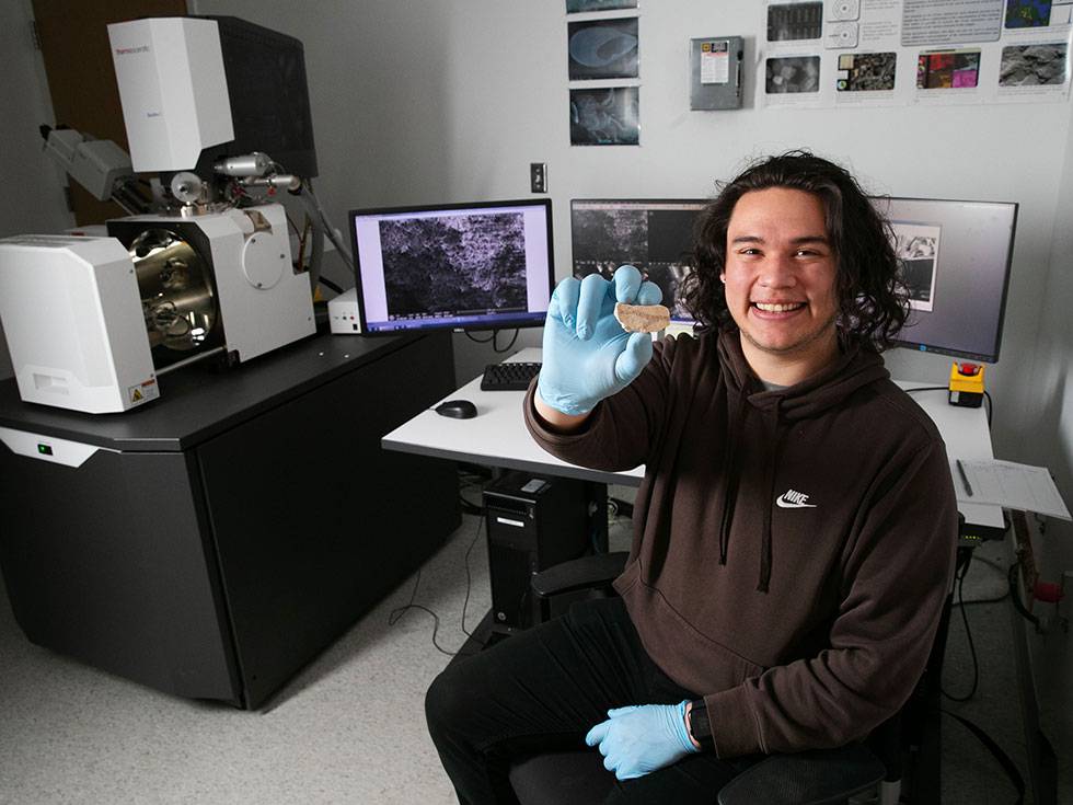
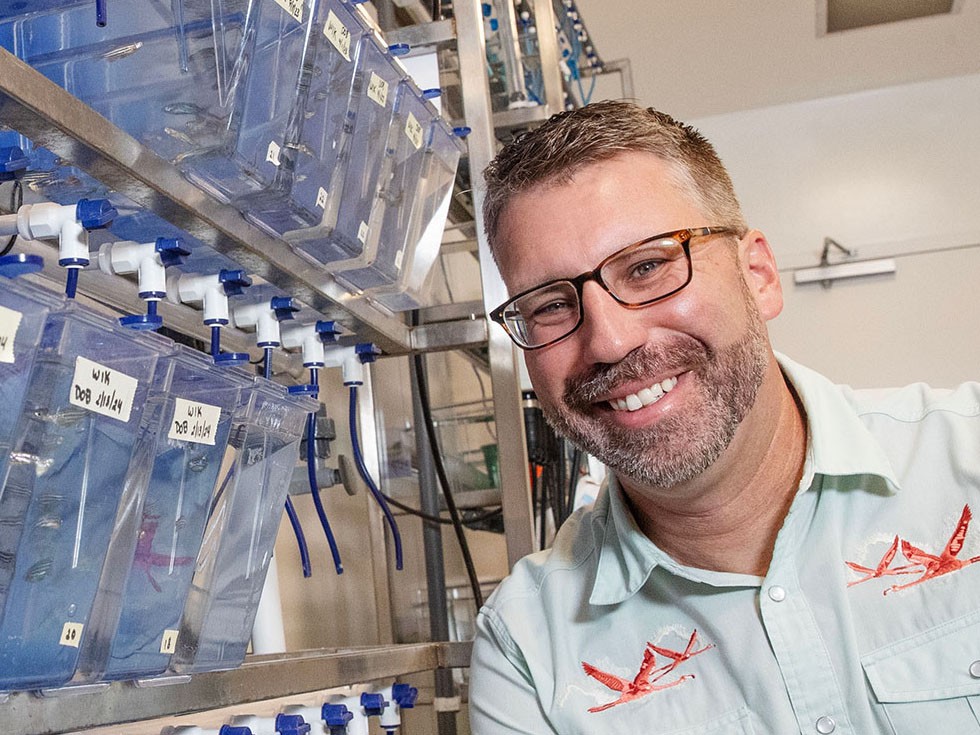
Hashimoto's Thyroiditis

Lindsey Wolf, Christina Campo, Nicole Sparbeck

Fall 2004

Home

This website is prepared for BI 348, an undergraduate college course in immunobiology.


What is Hashimoto's thyroidism?

Hashimoto's thyroiditis is a treatable autoimmune disease which attacks the thyroid resulting in inflammation and hypothyroidism. Hypothyroidism is the state where there is underproduction of thyroid hormone in the body.

Thyroid Importance

The thyroid is an endocrine gland responsible for:

•  Metabolic regulation

•  Thermal tolerance and regulation

•  Control of excitability & mood

•  Digestive function

•  Mental capacity

( http://www.nlm.nih.gov/medlineplus/ency/article/000371.htm 2004, October 8)

http://www.amwa-doc.org/index.cfm?objectId=943C5254-D567-0B25-51F593754A90B622

Prevalence

Named for the Japanese physician, Dr. Hakaru Hashimoto, who first described the condition in 1912, Hashimoto's Thyroiditis is the most prevalent form of hypothyroidism, affecting between 0.1-5% of all adults in Western countries, predominantly older women. In the United States alone, an estimated 17% of women are affected by age 60.

( http://www.endocrineweb.com/thyroiditis.html . 2004, October 8)


Mechanism

TH1 CD4+ (helper) T cells infiltrate the thyroid which leads to the production of autoantibodies against thyroperoxidase (TPO) and thyroglobulin (Tg). These antibodies against TPO and Tg in turn produce high levels of interferon- g (IFN- g ), a pro-inflammatory cytokine.


http://www.foxriverwatch.com/ thyroid.jpg.

As the disease progresses, there are fewer thyrocytes to secrete thyroglobulin. Less thyroglobulin yields less production of thyroxine (T 4 ). In order for the thyroid hormone to become activated, it must undergo monodeiodination in the liver to become triiodothyronine (T 3 ), the active form. If there are fewer thyrocytes secreting T 4, there will be less circulating active T 3.

http://www.endotext.org/adrenal/adrenal29/adrenal29.htm

A reduced [T 4 ] will signal the hypothalamus through negative feedback to produce more thyroid releasing hormone (TRH). TRH will then signal the anterior pituitary to release more thyroid stimulating hormone (TSH). TSH will attempt to stimulate the hormone to secrete more T 4. Continual hypersecretion of TSH and overstimulation of the thyroid will result in inflammation or goiter.

http://www.biol.andrews.edu/fb/spring/Chap.45-%20Endocrinology/4509.jpg 

Diagnosis:

Symptoms

Typical symptoms of Hashimoto's Thyroiditis include:

•  Intolerance to cold

•  Mild weight gain*

•  Fatigue*

•  Constipation

•  Enlarged neck or goiter

•  Dry skin

•  Hair loss or brittle hair*

•  Heavy menstrual cycles

•  Depression*

•  Difficulty concentrating or thinking*

The symptoms with asterisks are also characteristic of depression, and a definite diagnosis should be obtained through blood testing.

Laboratory Tests

Levels of serum TSH & free T 4 & T 3

•  TSH levels should be between 0.4-4.0 mIU/L

•  T 3 levels should be between 4.5-11.2 microgram/dl

•  T 4 levels should be between 100-200 ng/dl.


Presence of anti-thyroperoxidase and anti-thyroglobulin indicates Hashimoto's thyroiditis as unaffected individuals do not exhibit these antibodies.

http://www.clevelandclinicmeded.com/ccjm/april2003/zimmerman.htm

Thyroid Hormone Replacement

Because T 3 has a half-life of only 24 hours, synthetic T 4 in the form of levothyroxine (generic) must be taken through pill form once a day for the rest of the patient's life, so that it may be converted to T 3 . The most popular brand names of levothyroxine are Synthyroid™ and Levoxyl™. The dosage for the drug is 1.6 micrograms/ kilogram, or approximately 1.6 micrograms per 2.2 pounds.

  http://www.drharper.ca/thyroid_medications.htm

Potential Involvement of Fas and Its Ligand in the Pathogenesis of Hashimoto's Thyroiditis

Giordano et al. took it upon themselves to investigate the mechanism by which thyrocyte destruction occurs in Hashimoto's Thyroiditis (HT). The authors hypothesized that Fas and Fas-Ligand (FasL) may play a role in the death of thyrocytes in thyroid tissue. In order to test this hypothesis the authors performed several experiments to test for expression of Fas and FasL on HT thyrocytes as well as normal thyrocytes from non-toxic goiter (NTG) patients. The authors used mainly immunohistochemistry and flow cytometry to analyze their data and help to determine the involvement of Fas and FasL in autoimmune thyroid destruction.

Immunohistochemistry is a method by which specific proteins on cell surfaces can be detected using antibodies. For example, in this experiment the authors wanted to determine whether Fas was expressed on the surface of thyrocytes. To visualize this, the authors made a primary antibody to the Fas protein, and then made a secondary antibody which will bind to the primary antibody. The secondary antibody is labeled with a fluorescent tag which allows the location and presence of the particular protein to be seen under a microscope (American Heritage Dictionary, 2002).

In addition to immunohistochemistry, the authors employed the use of flow cytometry. This is another way to determine the presence of certain molecules on a cell's surface. Cells are passed through a beam of laser light and can be categorized by size and content. Antibodies specific to cell surface proteins are tagged with fluorescent dye and the amount of fluorescent light emitted can be counted and thereby the amount of labeled cell surface molecules can be measured (Parham, 2005).

Usually, the Fas-FasL interaction is used to dispose of unwanted lymphocytes during development and in the course of the immune response in order to prevent flooding of the immune system by unnecessary lymphocytes (Parham, 2005). The interaction of FasL on cytotoxic T-cell surfaces with Fas molecules expressed on the target cell surface causes the target cell to undergo apoptosis (Parham, 2005). Giordano et al. (1997) hypothesized that Fas and FasL may play a role in the pathogenesis of Hashimoto's Thyroiditis due to the massive amounts of apoptotic cell death in this disease.

In order to investigate the role of Fas and FasL the authors used two groups; HT thyrocytes and NTG thyrocytes. The first question the authors asked was whether or not there was a difference in Fas expression in HT thyrocytes versus NTG thyrocytes (Giordano et al., 1997). . Immunohistochemical and flow cytometric analysis of thyrocytes showed that HT thyrocytes express large amounts of Fas on their surface while NTG thyrocytes do not (Figure 1A).

The expression of Fas on HT thyrocytes may be a consequence of the initial inflammatory response that occurs in Hashimoto's Thyroiditis. The authors tested this by exposing NTG thyrocytes to various cytokines such as interleukin 1ß (IL-1ß), interferon-y (IFN-?) and tumor necrosis factor-a (TNF-a). The results showed that only IL-1ß was able to induce Fas expression in normal thyrocytes (Figure 2A). In addition, IL-1ß was abundantly present in HT tissue which makes the induction of Fas by IL-1 ß in HT a possibility (Figure 2E) (Giordano et al., 1997).

Although Fas was expressed in HT tissue, this does not mean that it can necessarily induce apoptosis in thyrocytes. In order to test the functionality of Fas, the authors induced Fas expression on normal thyrocytes with IL-1 ß, and then exposed Fas expressing thyrocytes to IL-1 ß again to trigger Fas. The authors found that Fas was able to cause apoptosis as evidence by the massive cell death observed in thyrocytes that had expressed Fas (Figure 3B).

In addition, the more IL-1 ß that was present, the more apoptosis occurred (Giordano et al, 1997). This has important implications for the progression of the disease and possibly slowing the progression by controlling the inflammatory response. Also, immunohistochemical analysis and the use of an in situ apoptosis detection kit showed that certain Fas positive thyrocytes in HT tissue were undergoing apoptosis while Fas negative NTG thyrocytes did not show signs of apoptosis (Giordano et al, 1997).

Although it seemed that Fas may be important in the pathogenesis of HT, the authors still had not investigated the role of FasL in thyrocyte destruction. Immunohistochemical analysis of frozen HT and normal thyrocytes as well as flow cytometric analysis of HT thyrocytes showed that FasL is expressed on both HT and NTG thyrocytes (Figure 4A).


Fig. 4. Constitutive expression of FasL by thyrocytes. ( A ) Immunohistochemical analysis of NTG, HT thyroid, or normal pancreatic (NP) cryostat sections exposed to control rabbit IgG or rabbit anti-FasL and visualized by immunoperoxidase. C ) RT-PCR in thyroid tissues. Lanes 1 and 2, samples of NTG tissues from two different individuals; lanes 3 and 4, samples from two different HT patients. phi  X174 DNA-Hae III digest was used as a size marker (M).

In addition, the authors performed reverse transcriptase polymerase chain reaction (RT-PCR) analysis to examine mRNA in both HT and NTG cells as well as infiltrating lymphocytes in the HT tissue. This type of analysis is useful for examining exactly what mRNA is expressed in cells at the time the mRNA is extracted. To do this, the mRNA is reverse transcribed into DNA by reverse transcriptase. After that, DNA polymerase creates a complimentary strand to produce double stranded DNA. This is the cDNA library. PCR is used to amplify the DNA so that there are enough copies to run on an agarose gel for electrophoresis ( Campbell , 2000). By doing this, the experimenters could see the mRNA that was being expressed at the time of extraction. The results showed that the expression of FasL by HT thyrocytes was four to five times greater than FasL expression by infiltrating lymphocytes (Figure 4C) (Giordano et al, 1997).

Once again, the authors wanted to know whether FasL was functional and tested functionality by observing the ability of normal thyrocytes to kill a lymphoma cells. They found that normal thyrocytes were able to kill Fas sensitive but not Fas insensitive lymphoma cells (Giordano et al, 1997). The blocking of Fas on lymphoma cells by a monoclonal antibody (mAB) prevented thyrocytes from killing lymphoma cells (Figure 5B).

Fig. 5. IL-1 beta -induced thyrocyte apoptosis induced by Fas-FasL interaction. ( B ) Apoptotic cell death in NTG thyrocytes exposed for 36 hours to IL-6 (200 U/ml), IL-1 beta (200 U/ml) plus control IgG1 (10 µg/ml), or IL-1 beta (200 U/ml) plus ZB4 mAb. Data represent the mean ± 1 SD of five different experiments. Cell viability was determined by orange acridine-ethidium bromide staining and fluorescence microscopy analysis.

This showed that FasL on thyrocytes is functional and able to induce apoptosis by interacting with function Fas (Giordano et al, 1997). In addition, since HT thyrocytes express large amounts of Fas as well as expressing FasL, the stimulation of these cells by IL-1 ß may induce apoptosis. The authors found that mABs that block Fas suppressed thyrocyte cell death even after being stimulated by IL-1 ß. This showed that IL-1 ß mediated induction of Fas on thyrocytes causes apoptotic cell death because they also express FasL (Giordano et al, 1997).

The results of this study seem to suggest a role for Fas and FasL in the progression of Hashimoto's Thyroiditis. Thyrocyte destruction can proceed independently of infiltrating T-lymphocytes following the inflammatory response. In addition, the progression of thyrocyte destruction may be accelerated due to the release of cytokine IL-1 ß by macrophages or monocytes during the course of the autoimmune response (Giordano et al, 1997). Because the expression of FasL on HT thyrocytes is much greater than that of infiltrating cytotoxic T cells, it seems that the cross-linking of HT thyrocytes may be the main source of thyrocyte destruction after the inflammatory response and cytotoxic T lymphocytes may not be directly involved in the destruction of thyrocytes (Giordano et al, 1997).


Caturegli, P., Hejazi, M., Suzuki, K., Dohan, O., Carrasco, N., Kohn, L. D., Rose, N. R. (2000) Hypothyroidism in transgenic mice expressing IFN- g in the thyroid. Proc. Natl. Acad. Sci. USA. 97, 1719-1724.

Due to the high prevalence of Hashimoto's thyroiditis and its role in causing hypothyroidism, it is important to understand the mechanisms responsible for the disease. One possible factor contributing to the susceptibility to autoimmune thyroiditis is the cytokine interferon- g (IFN- g ). Depending on the organ in which it is expressed, IFN- g has a number of normal functions in the immune system, including the expression of MHC class II genes in the thyroid gland. Past studies have implicated IFN- g with promoting thyroiditis, but the results were contradictory and inconclusive. The goal of this study was to determine whether chronic expression of IFN- g in the thyroid gland directly causes hypothyroidism.

Caturegli et al. conducted an in vivo experiment, using transgenic mice that expressed varying levels of IFN- g in the thyroid. They manipulated the expression levels by inserting IFN- g cDNA in between the thyroglobulin (TG) promoter and a human growth hormone gene (Fig. 6). Since the TG promoter supports transcription specifically in thyroid follicular cells, the manipulated DNA was able to express higher levels of IFN- g . Three lines of thyr- IFN- g transgenic mice were produced in this way, bearing either 22 (A), 18 (B), or 15 (C) copies of this transgene expressing IFN- g . These were compared to normal mice (N).

Fig. 6. Organization of thyr-IFN- g transgene.

They first determined the levels of IFN- g expression in each of the mouse strains. They found a significantly higher thyroidal expression of IFN- g in lines A and C transgenic mice than in line B transgenics (Fig. 7) ( P < 0.05 by two-way ANOVA). Also, by looking at IFN- g levels in various organs of the immune system, the researchers found that IFN- g was expressed almost solely in the thyroid. Then, to make sure the transgene was capable of producing functional IFN- g , they looked to see if the expressed IFN- g was capable of activating several classical IFN- g -responsive genes in thyroid cells. The three genes used code for MHC class I, MHC class II, and MHC class II transactivator (CIITA). The results indicated increased expression of all three of these genes in the thyroids of the transgenic mice (Fig. 8). Therefore, the IFN- g transgene was able to produce functional IFN- g . All these initial assessments were done through Northern blot analysis.

Fig. 7. Thyroidal expression of IFN-g in normal C57BL/6 mice (N) and the three transgenic mouse lines (A, B, and C), as assessed by Northern blot analysis.

Fig. 8. Ability of transgenic IFN-g to induce in the thyroid cells the expression of MHC class I, MHC class II, and CIITA genes in the three transgenic mouse lines (A, B, and C) and normal mice (N).

Once, the researchers had determined that their transgene was able to express functional IFN- g , primarily in the thyroid gland, and in a higher concentration in mouse lines A and C, they directed their efforts to understanding the effects of increased IFN- g expression in the thyroid. They found that the thyroid structure of the line A thyr -IFN- g transgenic mice was extensively disrupted. The typical thyroid follicle morphology was lost (Fig. 9A) and remaining cells were small and hypercellular (Fig. 9B, C). These morphological changes were compared to the thyroid architecture of normal mice (Fig. 9D). Additionally, the mutant mice developed hypothyroidism, the severity of which was directly proportional to the amount of IFN- g expressed in the thyroid. Therefore, the disease was more severe in line A and line C transgenic mice and not present in line B transgenics.

Fig. 9. Histological analysis of the thyroid glans in thyr-IFN-g transgenic and control mice. Hematoxylin/eosin staining of thyroid glands from line A transgenic mice at x10 magnification (A), x20 magnification (B), and x40 magnification (C). A normal thyroid from C57BL6 mouse is shown for comparison (D).

To determine the gene affected by IFN- g and responsible for the hypothyroidism, RNA probes were used to find any effects IFN- g had on the expression of thyroglobulin (TG), thyroperoxidase (TPO), TSH receptor (TSH-R), and sodium iodide symporter ( NIS ). Results indicated no effect on TG and little change in TPO and TSH-R, but a dramatic effect on NIS . The dark background for the NIS signal in Fig. 10 indicates the long exposure time needed to pick up the weakened signal in the transgenic mouse lines A and C. This shows the suppression of NIS expression in the mouse lines containing higher concentrations of IFN- g . However, when exposed to a lower concentration of IFN- g (as in line B transgenics), NIS transcription actually increased slightly. Additional in vitro experiments in other studies confirmed these results. Therefore, the almost complete suppression of NIS expression due to high levels of IFN- g may indicate its role in producing the classical features of Hashimoto's thyroiditis, with low iodine uptake and hypothyroidism. The authors explained the slight increase in NIS expression with low IFN- g concentrations by saying that, in the initial phase of the disease when IFN- g concentrations are still low, it is not uncommon to see a short-lived hyperthyroid phase involving increased iodine uptake. But as the disease progresses, IFN- g levels increase, NIS is suppressed, and iodine uptake decreases as hypothyroidism become apparent.

Fig. 10. Influence of transgenic IFN-g on the expression of thyroid-specific/ restricted genes.

However, the researchers wanted to make sure that these effects in the thyroid gland were a direct result of IFN- g expression rather than an indirect consequence of lymphocytes, which are also present and active in the thyroid. To do this, they crossed the thyr -IFN- g transgenic mice with RAG-2 knock-out mice, which lack functional B and T lymphocytes. They found that these mice, without lymphocytes, continue to display the previously described thyroid pathology. Additionally, they wanted to test to see if the IFN- g receptor was required for the observed thyroid changes. For this they crossed the thyr- IFN- g transgenic mice with IFN- g receptor knock-out mice and found that only the mice containing the IFN- g receptor developed the previously described thyroid pathology. This indicates that, to produce this thyroid phenotype, IFN- g has to bind to its plasma membrane receptor. Therefore, Caturegli et al.'s findings indicate that it is IFN- g expression in the thyroid gland that contributes to the suppression of NIS and, thus, the degradation of thyroid cells, decreased iodine uptake, and increased incidence of hypothyroidism.


Future Direction

In order to better understand the mechanisms behind human susceptibility to autoimmune thyroiditis it is important to continue research on various immune-modifying genes related to both general susceptibility to autoimmunity, including those that code for MHC molecules or cytokines like IFN- g , and thyroid-specific genes, like those for human thyroglobulin ( hTg ). This means that, in addition to quantitative data on the correlations between these genes and the presence of HT, there also need to be adequate characterization of the qualitative effects that result from these genes and their products, including their specific role in the development of autoimmune thyroiditis.

  REFERENCES

AMWA Facts about thyroid disease [Online]. Available: http://www.amwa-doc.org/index.cfm?objectId=943C5254-D567-0B25-51F593754A90B622 [2004, December 1].

Campbell (2000) RT-PCR Methodology. Department of Biology, Davidson College. [Online]. Available: http://www.bio.davidson.edu/courses/Immunology/Flash/RT_PCR.html. [2004. October 8].

Caturegli, P., Hejazi, M., Suzuki, K., Dohan, O., Carrasco, N., Kohn, L. D., Rose, N. R. (2000) Hypothyroidism in transgenic mice expressing IFN-? in the thyroid. Proc. Natl. Acad. Sci. USA. 97, 1719-1724.

Endocrine web: Thyroiditis [Online]. Available: http://www.endocrineweb.com/thyroiditis.html [2004, October 8].

Endotext.com - Adrenal Physiology And Diseases, Immune System, Effects on the Endocrine System [Online] Available: http://www.endotext.org/adrenal/adrenal29/adrenal29.htm. [2004, December 1].

Giordano, C., Stassi, G., de Maria, R., Todaro, M., Richiusa, P., Papoff, G., Ruberti, G., Bagnasco, M., Testi, R., Galluzzp, A. (1997) Potential involvement of Fas and its ligand in the pathogenesis of Hashimoto’s thyroiditis. Sci. 275, 960-963.

[Online] Available: http://www.biol.andrews.edu/fb/spring/Chap.45-%20Endocrinology/4509.jpg. [2004, December 1].

Medline: Medical Encyclopedia: Chronic thyroiditis (Hashimoto’s disease) [Online]. Available: http://www.nlm.nih.gov/medlineplus/ency/article/000371.htm [2004, October 8].

Parham, Peter. The Immune System. 2nd Ed. New York: Garland Science, 2005. p 348, 360.

The American Heritage® Stedman's Medical Dictionary (2002) Houghton Mifflin Company. Retrieved December 1, 2004 from www.dictionary.com

Thyroid Medications [Online] Available: http://www.drharper.ca/thyroid_medications.htm [2004, December 1]

What is the target TSH level in thyroid hormone replacement for primary hypothyroidism? [Online] Available: http://www.clevelandclinicmeded.com/ccjm/april2003/zimmerman.htm. [2004, December 1]

Contact Information:
c_campo@skidmore.edu
n_sparbe@skidmore.edu
l_wolf@skidmore.edu