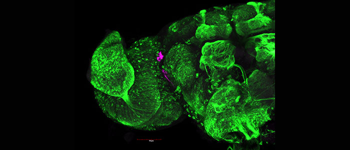
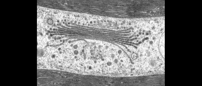
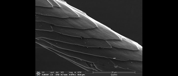
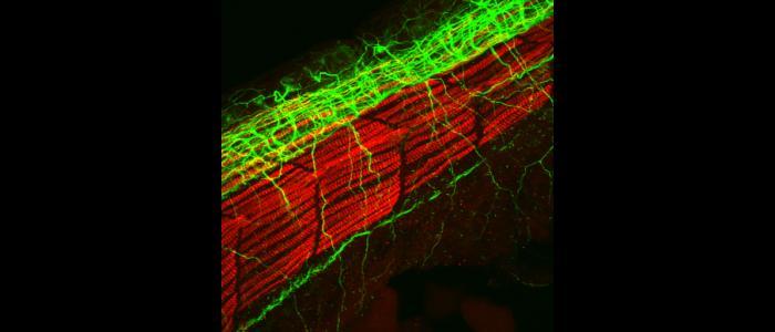
Skidmore McGraw Microscopy Imaging Center - SMMIC
The Skidmore McGraw Microscopy Imaging Center (SMMIC) was established in 2002 at Skidmore College. SMMIC is a core technology facility comprised of five modern laboratories in the Billie Tisch Center for Integrated Sciences (BTCIS) building that support light microscopy, confocal laser scanning microscopy (CLSM), scanning electron microscopy (SEM), transmission electron microscopy (TEM) and a variety of ancillary instruments used for the preparation of specimens. The mission of SMMIC is to promote and facilitate microscopy-based teaching and research at the college, enhance high-technology training in education, and provide user-friendly outreach to the surrounding communities. The equipments in SMMIC were acquired via fundings from the National Science Foundation Major Research Instrumentation program (NSF-MRI), the Lintilhac Foundation of Burlington, Vermont, the George I. Alden Trust of Massachusetts, several other private foundations and the college.
If you are a current member of Skidmore faculty or student and would like to know more about SMMIC, please contact Lily Kozel at 518-580-5088.
SMMIC is also open to external researchers or students from orther institutions or industry partners. For detail information, please contact Lily Kozel.
How to reserve instrument in SMMIC
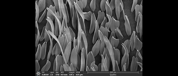
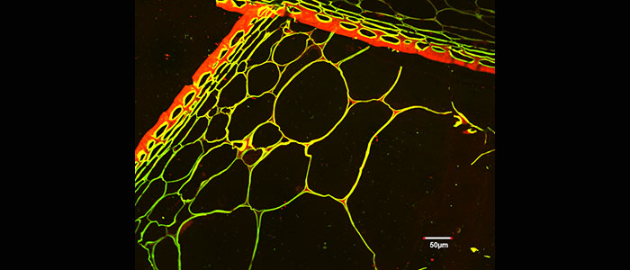
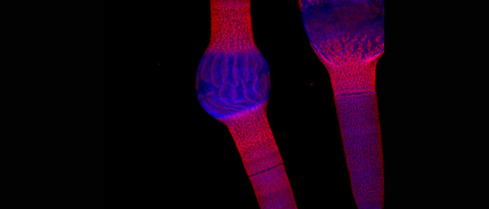
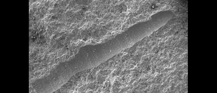
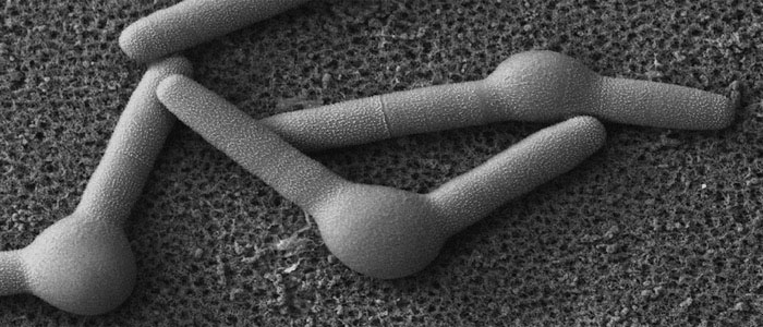
SMMIC Images on scientific journal covers

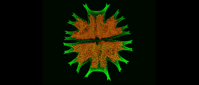
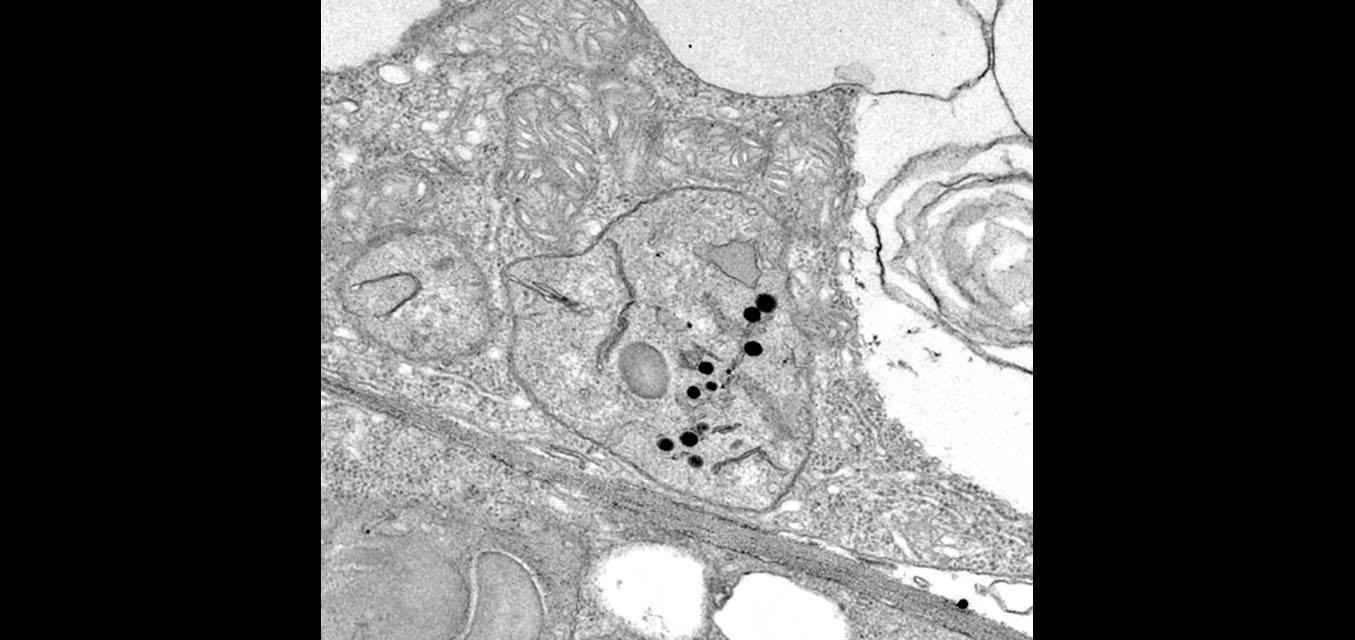
Published Journal Article: Josephine G LoRicco, et al. Journal of Experimental Botany, Volume 76, Issue 4, 25 February 2025, Pages 961–979. 3D Model of Penium margaritaceum cell reconstructed from the serial array tomography imaging on Scanning Electron Microscopy (SEM) in SMMIC. The internal features are colored as follows: chloroplasts (green), nuclei (cyan), Golgi apparatus (yellow), mitochondria (magenta), vacuoles (red), and cell wall septum (blue).