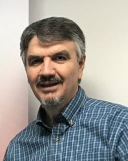
 Breno Leite
research scientist - sponsored research
Address:
Center for Integrated Sciences (CIS), Room 143
Department of Biology
Skidmore Microscopy Imaging Center (SMIC)
Department of Biology
Skidmore College
Saratoga Springs, NY 12866
Telephone: (518) 580-5073
Fax: (518) 580-5071
E-mail: bleite@skidmore.edu
Education:
- B.S., State University of Campinas - UNICAMP (Biology)
- M.S., State University of Campinas - UNICAMP (Genetics)
- Ph.D., Purdue University (Plant-Microbe Interactions)
- Postdoctoral Studies, University of Florida, University of Sao Paulo
Courses Taught:
- Biological Electron Microscopy (BI 311)
Research Interests:
Microscopy, microanalysis, cell-to-cell and cell-to-substrate interactions, biofilm architecture, cell wall composition, mucilaginous secretions, and low molecular weight metabolites functions.
Current Support:
- National Science Foundation grant to SMIC
Selected Publications:
- Leite B, Reddick KO, Boutaghou MN. 2021. Introducing a Novel, Extraction-free RT-PCR Kit that Simplifies and Accelerates Coronavirus Detection. Applications Paper – Shimadzu Scientific Instruments.
- Furlan, KP, Rebelo, DC, Leite B, Dias MVG, Klein, AN. 2017. Microstructural Characterization of Solid-State Reaction Phase Formed During Sintering of Hexagonal Boron Nitride with Iron. Microscopy and Microanalysis 23(5):1-6
- Leite B, Dziedzic R, Cruz, LF., Gillian-Daniel, AL., Nielsen, C. , De La Fuente, L. 2015. Microcontact Printing of Thiols: Changing the Way Cell Attachment is Investigated. Microscopy Today, 23(5): 24 - 29.
- Leite B, Dziedzic, R., Cruz LF., Gillian-Daniel AL., Nielsen C, De La Fuente L. 2015. The use of microcontact printing of thiols to evaluate attachment of Xylella fastidiosa Under Distinct Conditions of Calcium Availability. Microscopy and Microanalysis 21(S3):707-708
- Leite B & Andersen, PC. 2009. Localized accumulation of silicon (Si) in grape leaves affected by Pierce's disease. Microscopy and Microanalysis, 15 (S2), p. 918.
- Alves E., Leite B, Marucci, RC., Pascholati SF, Lopes JRS, Andersen P. C. 2008. Retention Sites for Xylella fastidiosa in Four Sharpshooter Vectors (Hemiptera: Cicadellidae) Analyzed by Scanning Electron Microscopy. Current Microbiology. 56:531-538.
- Ishida ML, Andersen, PC &, Leite B. 2004. Cecropin B antimicrobial activity against Xylella fastidiosa: effect on colony formation. Physiol. Molecular Plant Pathology. 64:73-81.
- Leite B, Andersen, PC. & Ishida ML. 2004. Colony Aggregation and biofilm formation in xylem chemistry-based media for Xylella fastidiosa. FEMS Microbiol. Lett. 230:283-290.
- Alves, E., Marucci, RC., Lopes, JRS. & Leite B. 2004. Relationship between the proportion of colonized vessels by Xylella fastidiosa in plum, coffee and citrus and the leaf symptomatology exhibited. J. Phytopathology. 152: 291-297.
- Schadeck, R.J.G., Randi, M.A.F., Buchi, D.F. & Leite B. 2002. Vacuolar system of ungerminated Colletotrichum Graminicola Conidia: convergence of Autophagic and endocytic pathways. FEMS Microbiol. Lett. 218: 277-283.
- Leite B, Ishida, ML, Alves, E, Carrer, H., Pascholati, SF, Kitajima EW. 2002. Genomics and X-ray microanalysis indicate that Ca++ and thiol mediate the Xylella fastidiosa aggregation and adhesion. Braz. J. of Med. and Biolog. Sci. 36:645-650.
Link for full list: https://scholar.google.com.au/citations?user=FU8_RLAAAAAJ&hl=en
Recent Abstracts/Presentations:
- Leite B, 2018. Sample Preparation for Microscopy (short course). Second at the Cerrado Microscopy Symposium – Federal University of Goias – Goiania - August 6, Goiania, Brazil
- Tosi A, Leite B, Ludka, IP, M Zucolotto M. 2014. Vicência meteorite found in Brazil: preliminary classification efforts based on traditional approaches and cathodoluminescence (CL) maps. Microscopy and Microanalysis, Hartford CT, August 2014
- Leite B, Bulloss, NA., 2010. Resolving EDS peak overlaps: how much resolution really matters? International Microscopy Conference (IMC 17), Rio de Janeiro, Brazil.
- Hack, G., Leite B, Guarrera, D, Hsia, RC. 2010. X-Ray Microanalysis of cylindrical structures emanating from dentinal tubules. Dental Research Conference. Washington, DC, USA.
- Leite B, Guarrera, D, Hsia, RC, Hack, G. 2010. Evidence that dentine cylindrical structures ("Tooth Worms") Are Involved in controlling calcium phosphate formation. Microscopy and Microanalysis, Portland, USA.
- Leite B. 2010. X- Ray Microanalysis: Reporting successful stories featuring solar cells and biological materials. FLAVS, Surface Analysis, Orlando, FL, USA.
Selected Awards:
- Scientific production featured on ResearchGate and Google citation index platforms
- Cover - Microscopy & Analysis Magazine catalog. 2010. International Microscopy Meeting 17 (IMC17). Rio de Janeiro, Brazil
- Research highlighted by the American Phytopathological Society website – Phakopsora pachyrhizi appressorium. 2007. USA
- Cover – Microscopy & Analysis Magazine: Puccinia paranahybae (Leite B. & EW Kitajima), 2000. USA
- Productivity award – CNPq-UFPR – Department of Biochemistry. 1997. Brazil
- Young Scientist Award – CNPq & Roberto Marinho Foundation. Brazil. Full Purdue University PhD program scholarship – Degree obtained in 1992
Memberships:
- American Phytopathological Society (APS)
- Microscopy Society of America (MSA)
- Purdue University Alumni Association
- Sigma Xi