
Facilities
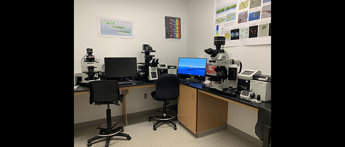
Most our experiments are performed in the Skidmore Microscopy Image Center (SMIC), a core technology facility composed of five modern laboratories in the Dana Science Complex that support light microscopy, confocal laser scanningmicroscopy (CLSM), scanning electron microscopy (SEM), transmission electron microscopy (TEM) and a variety of ancillary instruments used for the preparation of specimens.
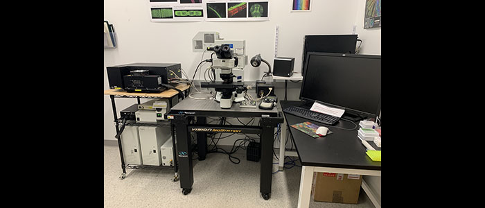
Lintilhac Light Microscopy Laboratory
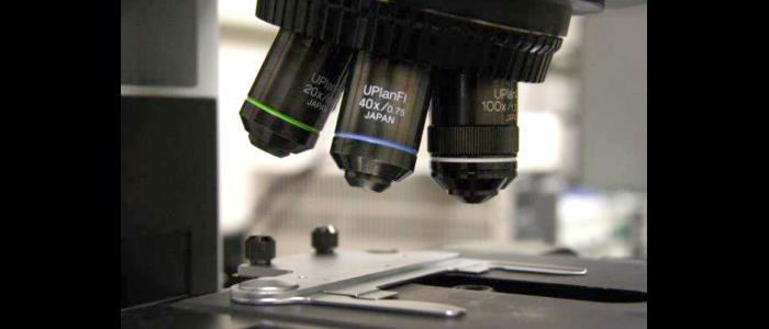
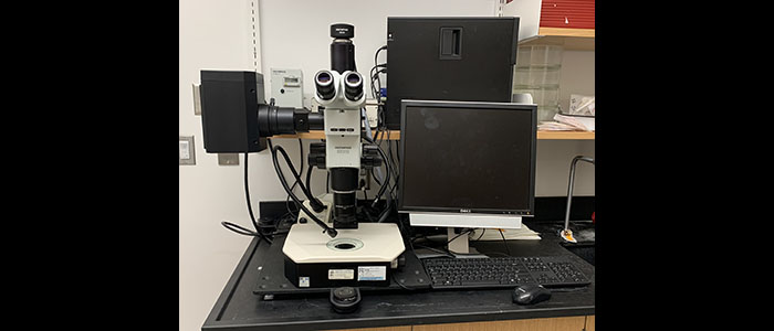
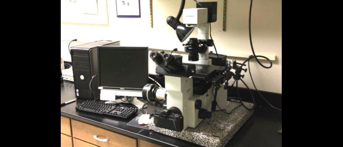
Our light microscopy laboratory contains three microscopy work stations:
-
Olympus BX63 light microscope with time-lapse function, with DIC and fluorescence optics (UV, blue and green filters), and an Olympus DP80 digital camera system.
-
Olympus IX83 inverted light microscope with time-lapse function, with DIC and fluorescence optics (UV, blue and green filters), and an Olympus DP80 digital camera system
-
Olympus BX-60 light microscope equipped with DIC and fluorescence optics and an Olympus DP73 digital camera for high-resolution imaging and video recording.
-
Olympus IX-70 inverted light microscope with phase and fluorescence optics and an Olympus DP70 digital camera for high-resolution imaging and video recording. This workstation is also equipped with a Narashige micromanipulator and microinjector for cell processing and injections.
-
Olympus SXZ-12 light microscope equipped with DIC and fluorescence optics and an Olympus DP73 digital camera for high-resolution imaging and video recording.
Confocal Laser Scanning Microscopy Laboratory
Olympus Fluoview 1000 confocal laser scanning microscope: This state-of-the-art confocal microscope is the latest addition to SMIC.
- Specifications
- Spectral and filter scan units
- five laser channels
- High-sensitivity GaAsP detector for imaging low-level fluorescence
- SIM scanner for simultaneous photostimulation during time-lapse imaging
- Enhanced, reliable objectives for live cell imaging
- High-precision focus and Z-tracking through extended time-lapse imaging
- 3D rendering and image analysis with advanced software
- Spectral unmixing for separating similar fluorophores
- Applications
- Multidimensional time-lapse imaging of live cells/organisms
- High-resolution 3D rendering of Z-stack images
- FRET and FRAP technologies
- Multi-laser combiner for simultaneous imaging of up to five different fluorophores
- DIC imaging
- Multi-point, tile and mapping scans
- 3D mosaic imaging
Scanning Electron Microscopy Laboratory
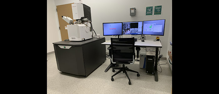
SPECIFICATIONS OF FEI QUATTRO S FESEM:
Illumination system: Field emission gun
Accelerating voltage: 0.2KV-30KV
Multi-purpose Sample Holder: simultaneous loading of 18 standard samples (ø 12 mm), three 45˚ pre-tilted samples, and two row bars (vertical, and 52˚ pre-tilted).
Detectors: conventional Everhart-Thornley SE detector with variable grid bias (high-Vacuum
mode)
low-vacuum mode gaseous SE detector
ESEM mode gaseous SE detector
Retractable Directional Back-scatter (DBS) Detector
Retractable STEM 3+ Detector
Modes: High-vacuum mode, low-vacuum mode, ESEM mode
Cooling Stage: temperature range from -20oC to 60oC.
X-ray Microanalysis: Thermo Scientific EDS UltraDry 60M (129 eV)
-Retractable UltraDry Premium EDS detector
-Active area of 60 mm2 and 129 eV energy resolution at Mn k-alpha
-Norvar window with proprietary evacuated tube
design for detection sensitivity to Be
-Analyzer electronics with up to 1,000,000 x-ray
input counts per second and 300,000 x-ray
output counts per second.
Transmission Electron Microscopy Laboratory
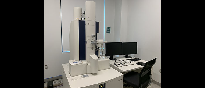
SPECIFICATIONS OF THE HITACHI HT7800 PREMIUM 120KV TEM:
|
Illumination System:
|
W or LaB6 Filament |
|
Accelerating Voltage:
|
120 kV maximum
|
| Magnification range: | x50 - x600,000 |
| Special features: |
Hitachi's unique "Dual Mode" compound objective lens provides both high contrast High resolution and advanced optimization of contrast; Eucentric high performance stage 5-axis goniometer for ultra-stable operation and Software and control panel driven z-height adjustment for efficient eucentric; Microprocessor-controlled, differential turbo molecular pump evacuation system to Advanced image navigation and storage of positions, tilt angles and orientations; Microtrace function indicating examined and unexamined areas of the specimen; EMIP software to automatically archive all saved images and imaging data for ease EMIP-3D tomography system and FBP reconstruction method |
| Sample holder: |
HT7800-MS2 Three Specimen One Touch Single Tilt Holder HT7800-SS Single Tilt Holder HT7800-RS Rotation Holder |
| Digital camera: |
AMT BioSprint16 High Definition Camera - 16 Megapixel Scientific CCD |
Ancillary Equipment
- Two ultramicrotomes: Reichert Ultracut E and Leica
- Sputter coater
- carbon coater
- Critical point dryer
- LKB knife maker
- Gene gun
Equipment is also available for freeze spraying, freeze slamming, freeze substitution and low-temperature plastic embedding.