
Penium research project images
Penium margaritaceum Is the model organism used in our laboratory. It is a unicellular desmid (Zygnematorphyceae; Virideplantae) that is commonly found in shallow freshwater wetlands.
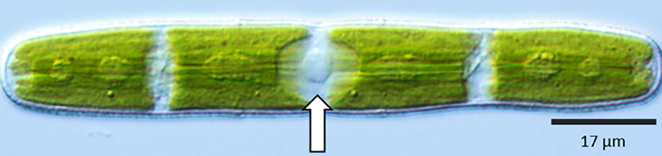
Penium ‘s nucleus (arrow) is located in the cell center, also known as the isthmus. 2-4 large chloroplasts surround the nucleus.
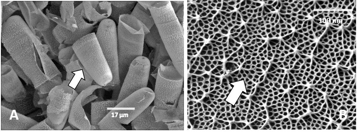
The cell wall of Penium is quite distinct. It contains many of the same polymers that are found in land plant cell walls. The walls can be easily isolated for biochemical and structural work (A arrows). The most notable feature of the cell wall is the outer layer that consists of a calcium-complexed pectin “lattice” (arrows, B). More information about the cell wall may be obtained at: doi: http:/ / dx. doi. org/ 10. 1104/ pp. 114. 236257 . A and B are SEM images.
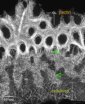
Transmission Electron Microscope (TEM) image of the Penium cell wall. The outer layer (OL) consists of tightly aggregated pectic homogalacturonan (HG) fibrils. The inner layer (IL) is composed of cellulose. The middle layer (ML) connects the outer and inner layers (arrows) and consists of pectic HG and rhamnogalacturonan-I.
The cell wall of Penium consists of polymers similar to those found in land plants.
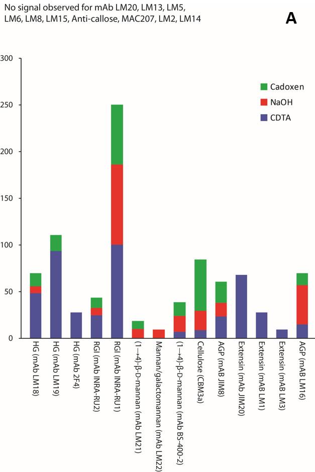
A is a summary diagram of a carbohydrate microarray analysis using antibodies with specificity to epitopes of land plant cell wall polysaccharides (courtesy of J. U. Fangel; see also DOI: 10.1111/j.1365-313X.2011.04686.x).
B The main component of the cell wall of Penium is pectin (homogalacturonan, HG, and rhamnogalacturonan-I, RG-I. The pectin fibrils are shown in this TEM micrograph.
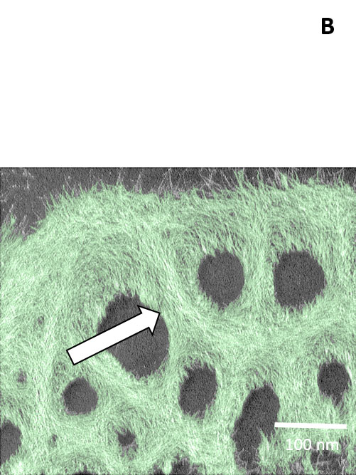
The outer layer of pectin in the Penium cell wall labeled with the monoclonal antibody, JIM5. Note the projections in a lattice-like arrangement (arrow).
The pectin “lattice” of the Penium cell wall as labeled with the monoclonal anti-body, JIM5 (arrow)
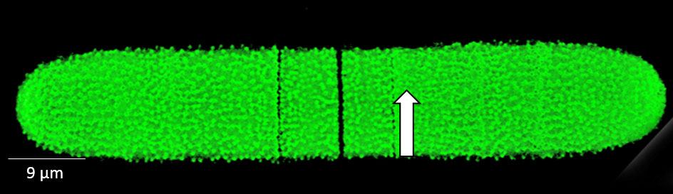
Penium secretes and incorporates new cell wall at its central isthmus zone (I). New wall material pushes older wall material toward the two poles (arrows). This cell was live labeled with the pectin-recognizing antibody JIM5, placed back in culture and allowed to grow. The dark zone represents new wall material.
The pectin “lattice” of the Penium cell wall as labeled with the monoclonal anti-body, JIM5 (arrow)
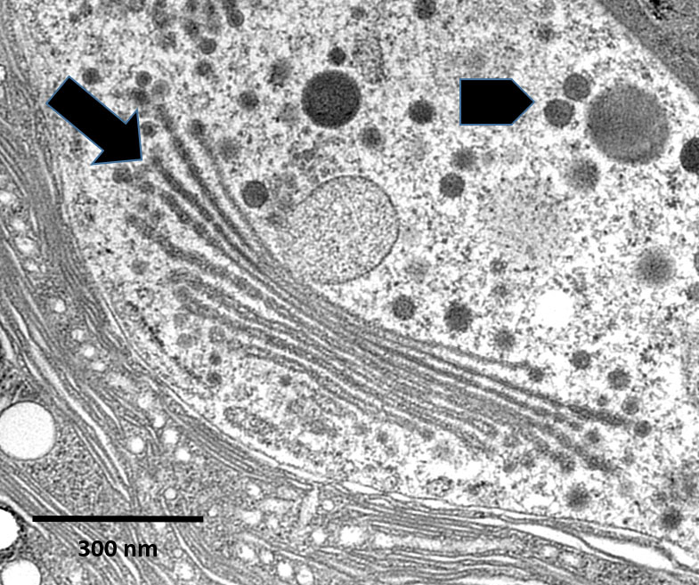
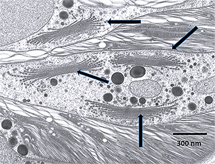
The Golgi body of Penium consists of 12-15 flattened cisternae (arrow). From the medial and trans faces of the Golgi body emerge various vesicles (arrowhead) carrying diverse materials to be secreted or sent to the vacuole. TEM micrograph
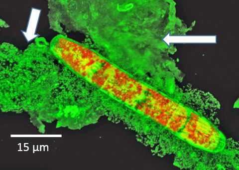
Confocal laser scanning microscope image of the extracellular polysaccharide secreted by Penium (arrows). This cell was labeled with an anti- extracellular polysaccharide antibody.
Confocal laser scanning microscope Image of the cytoplasmic channels of Penium (arrow; green fluorescence). This cell was labeled with the fluorescent dye, CFDA.
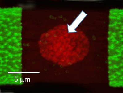
Confocal laser scanning microscope image of the nucleus of Penium (arrow). This cell was labeled with the nuclear dye, SYTO9.
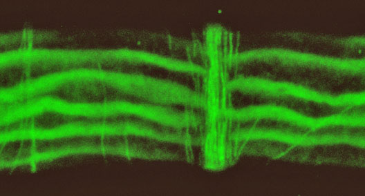
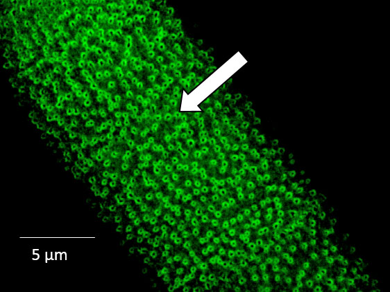
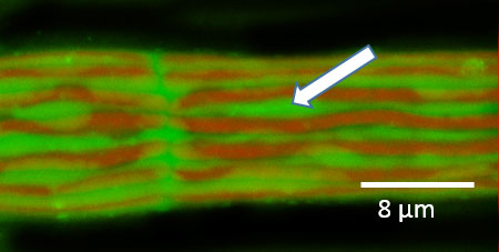
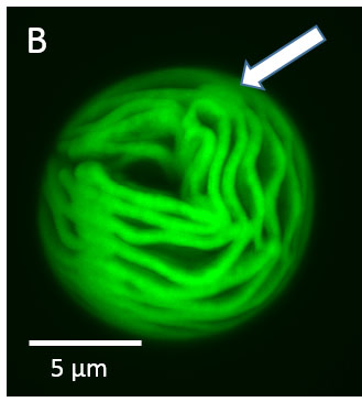
Confocal laser scanning microscope Image of the cortical microtubules (arrow). This cell was labeled with an anti-microtubule antibody.
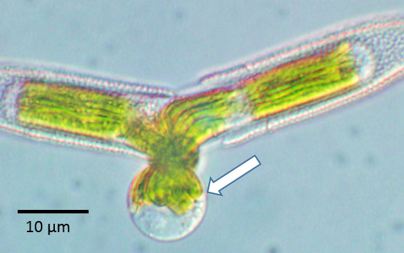
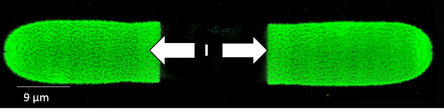
When cells placed in a mannitol-based buffer are treated with a cocktail of pectin- and cellulose-degrading enzymes, the protoplast emerges from the central isthmus zone (arrow).
Live protoplasts of Penium.
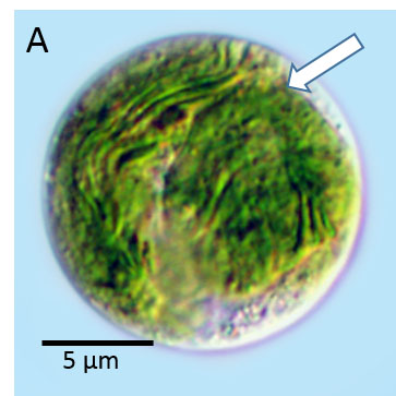
A is a Differential Interference Contrast or DIC- image of the protoplast (arrow).
B is a fluorescence image showing the autofluorescence of the chloroplast(arrow).